Calcium »
PDB 2eex-2f3z »
2exk »
Calcium in PDB 2exk: Structure of the FAMILY43 Beta-Xylosidase E187G From Geobacillus Stearothermophilus in Complex with Xylobiose
Enzymatic activity of Structure of the FAMILY43 Beta-Xylosidase E187G From Geobacillus Stearothermophilus in Complex with Xylobiose
All present enzymatic activity of Structure of the FAMILY43 Beta-Xylosidase E187G From Geobacillus Stearothermophilus in Complex with Xylobiose:
3.2.1.37;
3.2.1.37;
Protein crystallography data
The structure of Structure of the FAMILY43 Beta-Xylosidase E187G From Geobacillus Stearothermophilus in Complex with Xylobiose, PDB code: 2exk
was solved by
C.Brux,
K.Niefind,
D.Shallom-Shezifi,
Y.Shoham,
D.Schomburg,
with X-Ray Crystallography technique. A brief refinement statistics is given in the table below:
| Resolution Low / High (Å) | 2.23 / 2.20 |
| Space group | P 43 21 2 |
| Cell size a, b, c (Å), α, β, γ (°) | 139.870, 139.870, 232.025, 90.00, 90.00, 90.00 |
| R / Rfree (%) | 18.5 / 23.8 |
Calcium Binding Sites:
The binding sites of Calcium atom in the Structure of the FAMILY43 Beta-Xylosidase E187G From Geobacillus Stearothermophilus in Complex with Xylobiose
(pdb code 2exk). This binding sites where shown within
5.0 Angstroms radius around Calcium atom.
In total 4 binding sites of Calcium where determined in the Structure of the FAMILY43 Beta-Xylosidase E187G From Geobacillus Stearothermophilus in Complex with Xylobiose, PDB code: 2exk:
Jump to Calcium binding site number: 1; 2; 3; 4;
In total 4 binding sites of Calcium where determined in the Structure of the FAMILY43 Beta-Xylosidase E187G From Geobacillus Stearothermophilus in Complex with Xylobiose, PDB code: 2exk:
Jump to Calcium binding site number: 1; 2; 3; 4;
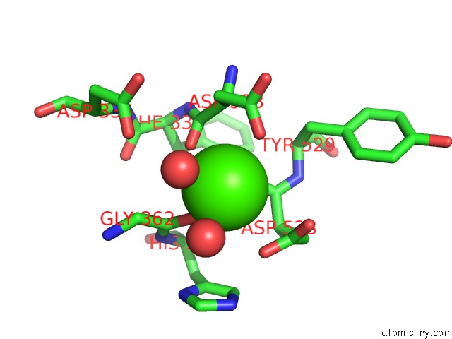
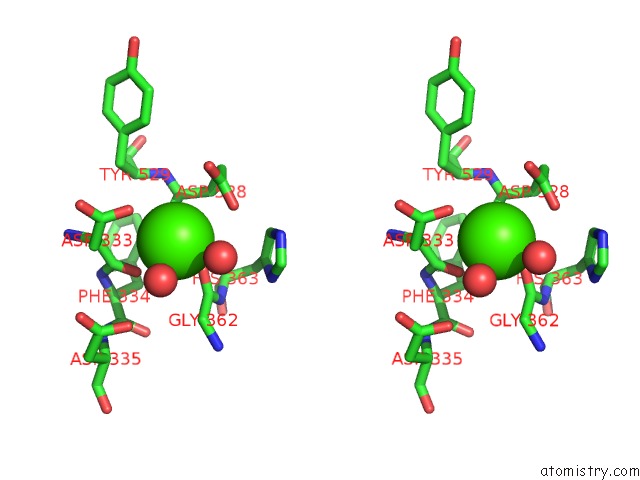
Calcium binding site 1 out of 4 in 2exk
Go back to
Calcium binding site 1 out
of 4 in the Structure of the FAMILY43 Beta-Xylosidase E187G From Geobacillus Stearothermophilus in Complex with Xylobiose
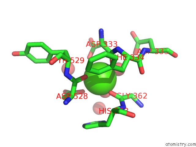
Mono view
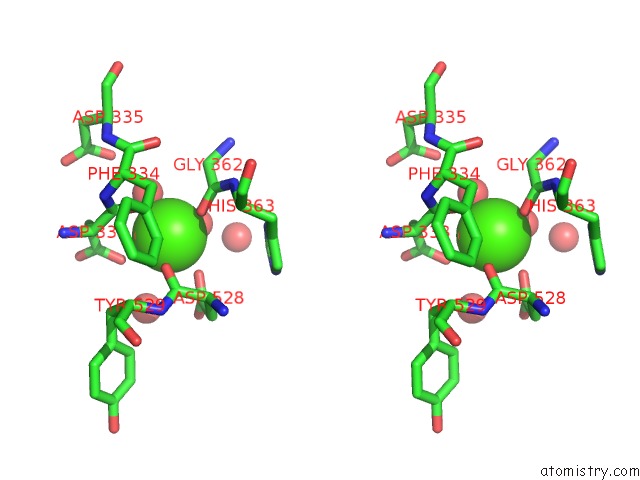
Stereo pair view
Mono view
Stereo pair view
A full contact list of Calcium with other atoms in the Ca binding
site number 1 of Structure of the FAMILY43 Beta-Xylosidase E187G From Geobacillus Stearothermophilus in Complex with Xylobiose within 5.0Å range:
|
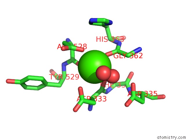
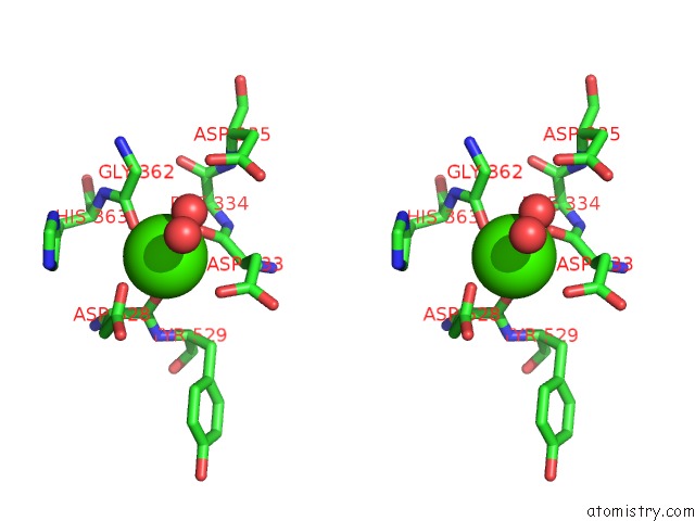
Calcium binding site 2 out of 4 in 2exk
Go back to
Calcium binding site 2 out
of 4 in the Structure of the FAMILY43 Beta-Xylosidase E187G From Geobacillus Stearothermophilus in Complex with Xylobiose
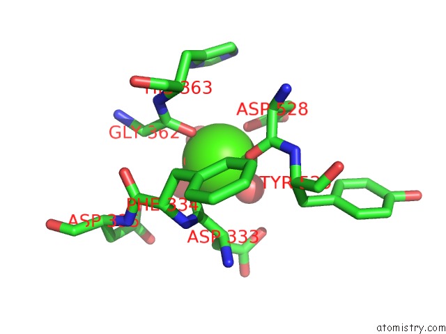
Mono view
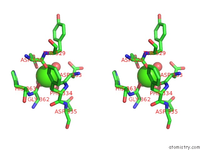
Stereo pair view
Mono view
Stereo pair view
A full contact list of Calcium with other atoms in the Ca binding
site number 2 of Structure of the FAMILY43 Beta-Xylosidase E187G From Geobacillus Stearothermophilus in Complex with Xylobiose within 5.0Å range:
|
Calcium binding site 3 out of 4 in 2exk
Go back to
Calcium binding site 3 out
of 4 in the Structure of the FAMILY43 Beta-Xylosidase E187G From Geobacillus Stearothermophilus in Complex with Xylobiose
Mono view
Stereo pair view
Mono view
Stereo pair view
A full contact list of Calcium with other atoms in the Ca binding
site number 3 of Structure of the FAMILY43 Beta-Xylosidase E187G From Geobacillus Stearothermophilus in Complex with Xylobiose within 5.0Å range:
|
Calcium binding site 4 out of 4 in 2exk
Go back to
Calcium binding site 4 out
of 4 in the Structure of the FAMILY43 Beta-Xylosidase E187G From Geobacillus Stearothermophilus in Complex with Xylobiose
Mono view
Stereo pair view
Mono view
Stereo pair view
A full contact list of Calcium with other atoms in the Ca binding
site number 4 of Structure of the FAMILY43 Beta-Xylosidase E187G From Geobacillus Stearothermophilus in Complex with Xylobiose within 5.0Å range:
|
Reference:
C.Brux,
A.Ben-David,
D.Shallom-Shezifi,
M.Leon,
K.Niefind,
G.Shoham,
Y.Shoham,
D.Schomburg.
The Structure of An Inverting GH43 Beta-Xylosidase From Geobacillus Stearothermophilus with Its Substrate Reveals the Role of the Three Catalytic Residues. J.Mol.Biol. V. 359 97 2006.
ISSN: ISSN 0022-2836
PubMed: 16631196
DOI: 10.1016/J.JMB.2006.03.005
Page generated: Tue Jul 8 05:25:33 2025
ISSN: ISSN 0022-2836
PubMed: 16631196
DOI: 10.1016/J.JMB.2006.03.005
Last articles
Cl in 8ASFCl in 8ASY
Cl in 8ATP
Cl in 8ATE
Cl in 8AQ6
Cl in 8AQI
Cl in 8ASE
Cl in 8ARZ
Cl in 8ARY
Cl in 8ARX