Calcium »
PDB 2f59-2foc »
2fh6 »
Calcium in PDB 2fh6: Crystal Structure Analysis of Klebsiella Pneumoniae Pullulanase Complexed with Glucose
Enzymatic activity of Crystal Structure Analysis of Klebsiella Pneumoniae Pullulanase Complexed with Glucose
All present enzymatic activity of Crystal Structure Analysis of Klebsiella Pneumoniae Pullulanase Complexed with Glucose:
3.2.1.41;
3.2.1.41;
Protein crystallography data
The structure of Crystal Structure Analysis of Klebsiella Pneumoniae Pullulanase Complexed with Glucose, PDB code: 2fh6
was solved by
B.Mikami,
H.Iwamoto,
Y.Katsuya,
H.-J.Yoon,
E.Demirkan-Sarikaya,
D.Malle,
with X-Ray Crystallography technique. A brief refinement statistics is given in the table below:
| Resolution Low / High (Å) | 14.96 / 1.80 |
| Space group | C 1 2 1 |
| Cell size a, b, c (Å), α, β, γ (°) | 149.263, 60.420, 133.650, 90.00, 111.98, 90.00 |
| R / Rfree (%) | 18.5 / 21.2 |
Calcium Binding Sites:
The binding sites of Calcium atom in the Crystal Structure Analysis of Klebsiella Pneumoniae Pullulanase Complexed with Glucose
(pdb code 2fh6). This binding sites where shown within
5.0 Angstroms radius around Calcium atom.
In total 4 binding sites of Calcium where determined in the Crystal Structure Analysis of Klebsiella Pneumoniae Pullulanase Complexed with Glucose, PDB code: 2fh6:
Jump to Calcium binding site number: 1; 2; 3; 4;
In total 4 binding sites of Calcium where determined in the Crystal Structure Analysis of Klebsiella Pneumoniae Pullulanase Complexed with Glucose, PDB code: 2fh6:
Jump to Calcium binding site number: 1; 2; 3; 4;
Calcium binding site 1 out of 4 in 2fh6
Go back to
Calcium binding site 1 out
of 4 in the Crystal Structure Analysis of Klebsiella Pneumoniae Pullulanase Complexed with Glucose

Mono view
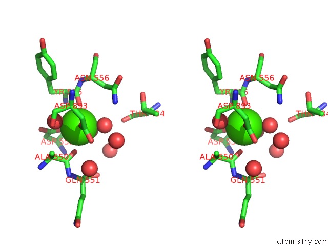
Stereo pair view
Mono view
Stereo pair view
A full contact list of Calcium with other atoms in the Ca binding
site number 1 of Crystal Structure Analysis of Klebsiella Pneumoniae Pullulanase Complexed with Glucose within 5.0Å range:
|
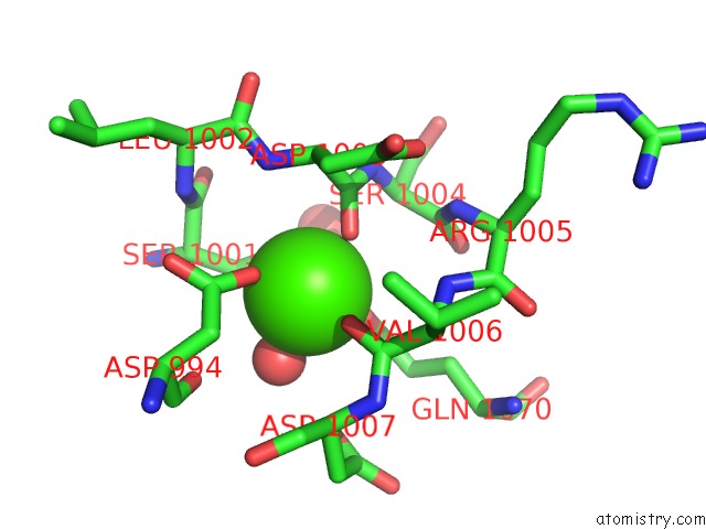
Calcium binding site 2 out of 4 in 2fh6
Go back to
Calcium binding site 2 out
of 4 in the Crystal Structure Analysis of Klebsiella Pneumoniae Pullulanase Complexed with Glucose
Mono view

Stereo pair view
Mono view
Stereo pair view
A full contact list of Calcium with other atoms in the Ca binding
site number 2 of Crystal Structure Analysis of Klebsiella Pneumoniae Pullulanase Complexed with Glucose within 5.0Å range:
|
Calcium binding site 3 out of 4 in 2fh6
Go back to
Calcium binding site 3 out
of 4 in the Crystal Structure Analysis of Klebsiella Pneumoniae Pullulanase Complexed with Glucose
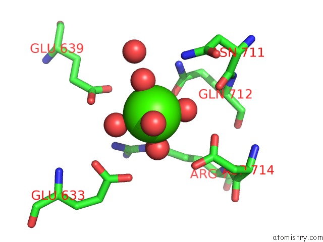
Mono view
Stereo pair view
Mono view
Stereo pair view
A full contact list of Calcium with other atoms in the Ca binding
site number 3 of Crystal Structure Analysis of Klebsiella Pneumoniae Pullulanase Complexed with Glucose within 5.0Å range:
|
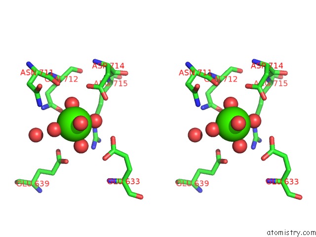
Calcium binding site 4 out of 4 in 2fh6
Go back to
Calcium binding site 4 out
of 4 in the Crystal Structure Analysis of Klebsiella Pneumoniae Pullulanase Complexed with Glucose

Mono view

Stereo pair view
Mono view
Stereo pair view
A full contact list of Calcium with other atoms in the Ca binding
site number 4 of Crystal Structure Analysis of Klebsiella Pneumoniae Pullulanase Complexed with Glucose within 5.0Å range:
|
Reference:
B.Mikami,
H.Iwamoto,
D.Malle,
H.-J.Yoon,
E.Demirkan-Sarikaya,
Y.Mezaki,
Y.Katsuya.
Crystal Structure of Pullulanase: Evidence For Parallel Binding of Oligosaccharides in the Active Site J.Mol.Biol. V. 359 690 2006.
ISSN: ISSN 0022-2836
PubMed: 16650854
DOI: 10.1016/J.JMB.2006.03.058
Page generated: Fri Jul 12 10:27:12 2024
ISSN: ISSN 0022-2836
PubMed: 16650854
DOI: 10.1016/J.JMB.2006.03.058
Last articles
Zn in 9MJ5Zn in 9HNW
Zn in 9G0L
Zn in 9FNE
Zn in 9DZN
Zn in 9E0I
Zn in 9D32
Zn in 9DAK
Zn in 8ZXC
Zn in 8ZUF