Calcium »
PDB 2fod-2g81 »
2ftl »
Calcium in PDB 2ftl: Crystal Structure of Trypsin Complexed with Bpti at 100K
Enzymatic activity of Crystal Structure of Trypsin Complexed with Bpti at 100K
All present enzymatic activity of Crystal Structure of Trypsin Complexed with Bpti at 100K:
3.4.21.4;
3.4.21.4;
Protein crystallography data
The structure of Crystal Structure of Trypsin Complexed with Bpti at 100K, PDB code: 2ftl
was solved by
W.M.Hanson,
M.P.Horvath,
D.P.Goldenberg,
with X-Ray Crystallography technique. A brief refinement statistics is given in the table below:
| Resolution Low / High (Å) | 20.00 / 1.62 |
| Space group | I 2 2 2 |
| Cell size a, b, c (Å), α, β, γ (°) | 74.726, 81.906, 124.261, 90.00, 90.00, 90.00 |
| R / Rfree (%) | 21.4 / 22.7 |
Other elements in 2ftl:
The structure of Crystal Structure of Trypsin Complexed with Bpti at 100K also contains other interesting chemical elements:
| Sodium | (Na) | 1 atom |
Calcium Binding Sites:
The binding sites of Calcium atom in the Crystal Structure of Trypsin Complexed with Bpti at 100K
(pdb code 2ftl). This binding sites where shown within
5.0 Angstroms radius around Calcium atom.
In total 2 binding sites of Calcium where determined in the Crystal Structure of Trypsin Complexed with Bpti at 100K, PDB code: 2ftl:
Jump to Calcium binding site number: 1; 2;
In total 2 binding sites of Calcium where determined in the Crystal Structure of Trypsin Complexed with Bpti at 100K, PDB code: 2ftl:
Jump to Calcium binding site number: 1; 2;
Calcium binding site 1 out of 2 in 2ftl
Go back to
Calcium binding site 1 out
of 2 in the Crystal Structure of Trypsin Complexed with Bpti at 100K
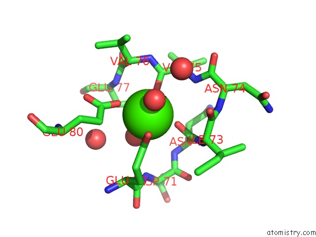
Mono view
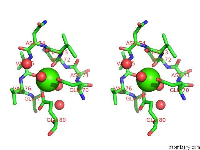
Stereo pair view
Mono view
Stereo pair view
A full contact list of Calcium with other atoms in the Ca binding
site number 1 of Crystal Structure of Trypsin Complexed with Bpti at 100K within 5.0Å range:
|
Calcium binding site 2 out of 2 in 2ftl
Go back to
Calcium binding site 2 out
of 2 in the Crystal Structure of Trypsin Complexed with Bpti at 100K
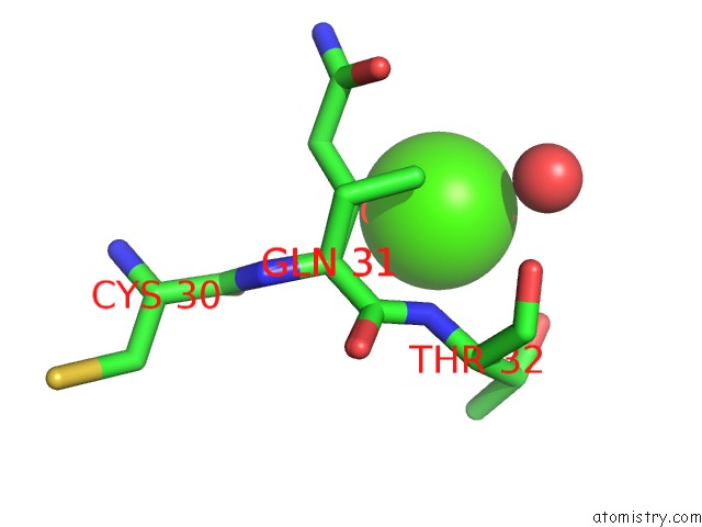
Mono view
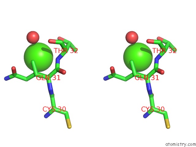
Stereo pair view
Mono view
Stereo pair view
A full contact list of Calcium with other atoms in the Ca binding
site number 2 of Crystal Structure of Trypsin Complexed with Bpti at 100K within 5.0Å range:
|
Reference:
W.M.Hanson,
G.J.Domek,
M.P.Horvath,
D.P.Goldenberg.
Rigidification of A Flexible Protease Inhibitor Variant Upon Binding to Trypsin. J.Mol.Biol. V. 366 230 2007.
ISSN: ISSN 0022-2836
PubMed: 17157870
DOI: 10.1016/J.JMB.2006.11.003
Page generated: Fri Jul 12 10:36:57 2024
ISSN: ISSN 0022-2836
PubMed: 17157870
DOI: 10.1016/J.JMB.2006.11.003
Last articles
Zn in 9JYWZn in 9IR4
Zn in 9IR3
Zn in 9GMX
Zn in 9GMW
Zn in 9JEJ
Zn in 9ERF
Zn in 9ERE
Zn in 9EGV
Zn in 9EGW