Calcium »
PDB 2fod-2g81 »
2fvy »
Calcium in PDB 2fvy: High Resolution Glucose Bound Crystal Structure of Ggbp
Protein crystallography data
The structure of High Resolution Glucose Bound Crystal Structure of Ggbp, PDB code: 2fvy
was solved by
M.J.Borrok,
L.L.Kiessling,
K.T.Forest,
with X-Ray Crystallography technique. A brief refinement statistics is given in the table below:
| Resolution Low / High (Å) | 9.00 / 0.92 |
| Space group | C 1 2 1 |
| Cell size a, b, c (Å), α, β, γ (°) | 119.989, 36.238, 80.101, 90.00, 124.50, 90.00 |
| R / Rfree (%) | 10.9 / 13 |
Calcium Binding Sites:
The binding sites of Calcium atom in the High Resolution Glucose Bound Crystal Structure of Ggbp
(pdb code 2fvy). This binding sites where shown within
5.0 Angstroms radius around Calcium atom.
In total only one binding site of Calcium was determined in the High Resolution Glucose Bound Crystal Structure of Ggbp, PDB code: 2fvy:
In total only one binding site of Calcium was determined in the High Resolution Glucose Bound Crystal Structure of Ggbp, PDB code: 2fvy:
Calcium binding site 1 out of 1 in 2fvy
Go back to
Calcium binding site 1 out
of 1 in the High Resolution Glucose Bound Crystal Structure of Ggbp

Mono view
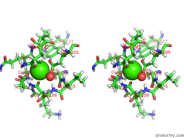
Stereo pair view
Mono view
Stereo pair view
A full contact list of Calcium with other atoms in the Ca binding
site number 1 of High Resolution Glucose Bound Crystal Structure of Ggbp within 5.0Å range:
|
Reference:
M.J.Borrok,
L.L.Kiessling,
K.T.Forest.
Conformational Changes of Glucose/Galactose-Binding Protein Illuminated By Open, Unliganded, and Ultra-High-Resolution Ligand-Bound Structures. Protein Sci. V. 16 1032 2007.
ISSN: ISSN 0961-8368
PubMed: 17473016
DOI: 10.1110/PS.062707807
Page generated: Tue Jul 8 05:38:15 2025
ISSN: ISSN 0961-8368
PubMed: 17473016
DOI: 10.1110/PS.062707807
Last articles
Cl in 5G6QCl in 5G6P
Cl in 5G6O
Cl in 5G6L
Cl in 5G6N
Cl in 5G6M
Cl in 5G6K
Cl in 5G6I
Cl in 5G6J
Cl in 5G6H