Calcium »
PDB 2fod-2g81 »
2g0y »
Calcium in PDB 2g0y: Crystal Structure of A Lumenal Pentapeptide Repeat Protein From Cyanothece Sp 51142 at 2.3 Angstrom Resolution. Tetragonal Crystal Form
Protein crystallography data
The structure of Crystal Structure of A Lumenal Pentapeptide Repeat Protein From Cyanothece Sp 51142 at 2.3 Angstrom Resolution. Tetragonal Crystal Form, PDB code: 2g0y
was solved by
M.A.Kennedy,
S.Ni,
G.W.Buchko,
H.Robinson,
with X-Ray Crystallography technique. A brief refinement statistics is given in the table below:
| Resolution Low / High (Å) | 500.00 / 2.30 |
| Space group | I 41 2 2 |
| Cell size a, b, c (Å), α, β, γ (°) | 86.614, 86.614, 89.745, 90.00, 90.00, 90.00 |
| R / Rfree (%) | 23.3 / 26.6 |
Calcium Binding Sites:
The binding sites of Calcium atom in the Crystal Structure of A Lumenal Pentapeptide Repeat Protein From Cyanothece Sp 51142 at 2.3 Angstrom Resolution. Tetragonal Crystal Form
(pdb code 2g0y). This binding sites where shown within
5.0 Angstroms radius around Calcium atom.
In total 3 binding sites of Calcium where determined in the Crystal Structure of A Lumenal Pentapeptide Repeat Protein From Cyanothece Sp 51142 at 2.3 Angstrom Resolution. Tetragonal Crystal Form, PDB code: 2g0y:
Jump to Calcium binding site number: 1; 2; 3;
In total 3 binding sites of Calcium where determined in the Crystal Structure of A Lumenal Pentapeptide Repeat Protein From Cyanothece Sp 51142 at 2.3 Angstrom Resolution. Tetragonal Crystal Form, PDB code: 2g0y:
Jump to Calcium binding site number: 1; 2; 3;
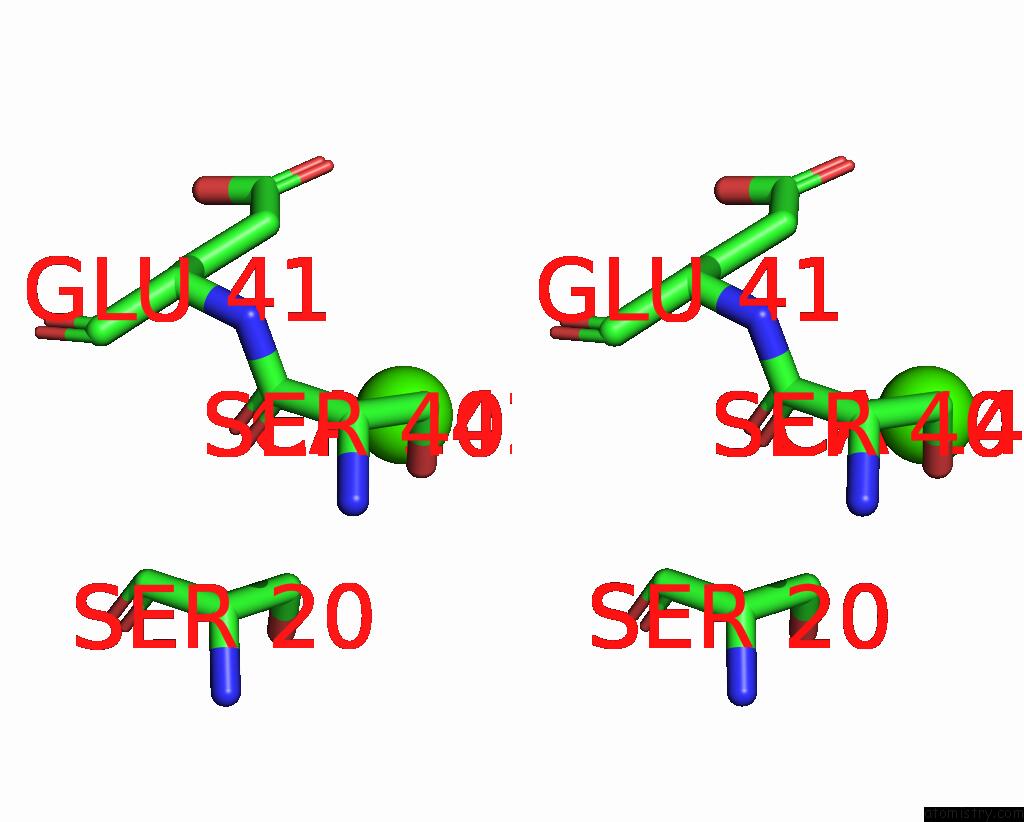
Calcium binding site 1 out of 3 in 2g0y
Go back to
Calcium binding site 1 out
of 3 in the Crystal Structure of A Lumenal Pentapeptide Repeat Protein From Cyanothece Sp 51142 at 2.3 Angstrom Resolution. Tetragonal Crystal Form
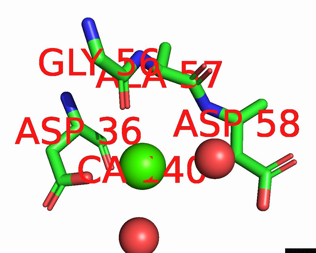
Mono view
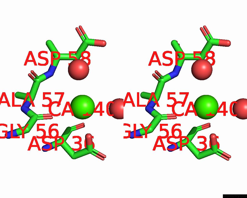
Stereo pair view
Mono view
Stereo pair view
A full contact list of Calcium with other atoms in the Ca binding
site number 1 of Crystal Structure of A Lumenal Pentapeptide Repeat Protein From Cyanothece Sp 51142 at 2.3 Angstrom Resolution. Tetragonal Crystal Form within 5.0Å range:
|
Calcium binding site 2 out of 3 in 2g0y
Go back to
Calcium binding site 2 out
of 3 in the Crystal Structure of A Lumenal Pentapeptide Repeat Protein From Cyanothece Sp 51142 at 2.3 Angstrom Resolution. Tetragonal Crystal Form
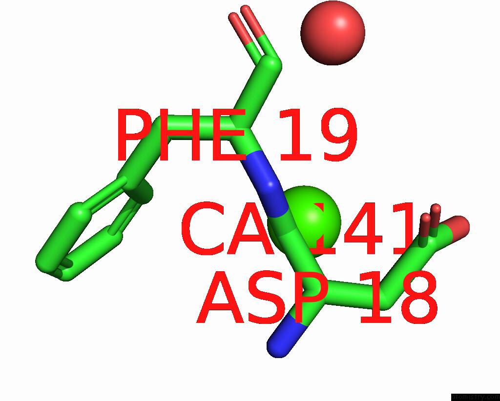
Mono view
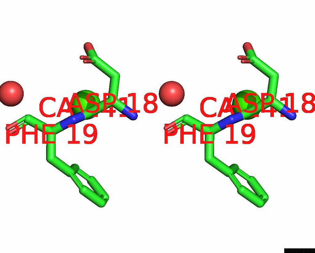
Stereo pair view
Mono view
Stereo pair view
A full contact list of Calcium with other atoms in the Ca binding
site number 2 of Crystal Structure of A Lumenal Pentapeptide Repeat Protein From Cyanothece Sp 51142 at 2.3 Angstrom Resolution. Tetragonal Crystal Form within 5.0Å range:
|
Calcium binding site 3 out of 3 in 2g0y
Go back to
Calcium binding site 3 out
of 3 in the Crystal Structure of A Lumenal Pentapeptide Repeat Protein From Cyanothece Sp 51142 at 2.3 Angstrom Resolution. Tetragonal Crystal Form

Mono view
Stereo pair view
Mono view
Stereo pair view
A full contact list of Calcium with other atoms in the Ca binding
site number 3 of Crystal Structure of A Lumenal Pentapeptide Repeat Protein From Cyanothece Sp 51142 at 2.3 Angstrom Resolution. Tetragonal Crystal Form within 5.0Å range:
|
Reference:
G.W.Buchko,
S.Ni,
H.Robinson,
E.A.Welsh,
H.B.Pakrasi,
M.A.Kennedy.
Characterization of Two Potentially Universal Turn Motifs That Shape the Repeated Five-Residues Fold - Crystal Structure of A Lumenal Pentapeptide Repeat Protein From Cyanothece 51142 Protein Sci. V. 15 2579 2006.
ISSN: ISSN 0961-8368
PubMed: 17075135
DOI: 10.1110/PS.062407506
Page generated: Fri Jul 12 10:40:30 2024
ISSN: ISSN 0961-8368
PubMed: 17075135
DOI: 10.1110/PS.062407506
Last articles
Zn in 9MJ5Zn in 9HNW
Zn in 9G0L
Zn in 9FNE
Zn in 9DZN
Zn in 9E0I
Zn in 9D32
Zn in 9DAK
Zn in 8ZXC
Zn in 8ZUF