Calcium »
PDB 2fod-2g81 »
2g55 »
Calcium in PDB 2g55: Anomalous Substructure of Trypsin (P3121)
Enzymatic activity of Anomalous Substructure of Trypsin (P3121)
All present enzymatic activity of Anomalous Substructure of Trypsin (P3121):
3.4.21.4;
3.4.21.4;
Protein crystallography data
The structure of Anomalous Substructure of Trypsin (P3121), PDB code: 2g55
was solved by
C.Mueller-Dieckmann,
M.S.Weiss,
with X-Ray Crystallography technique. A brief refinement statistics is given in the table below:
| Resolution Low / High (Å) | 30.00 / 1.82 |
| Space group | P 31 2 1 |
| Cell size a, b, c (Å), α, β, γ (°) | 54.590, 54.590, 107.090, 90.00, 90.00, 120.00 |
| R / Rfree (%) | 15.8 / 20.8 |
Other elements in 2g55:
The structure of Anomalous Substructure of Trypsin (P3121) also contains other interesting chemical elements:
| Chlorine | (Cl) | 3 atoms |
Calcium Binding Sites:
The binding sites of Calcium atom in the Anomalous Substructure of Trypsin (P3121)
(pdb code 2g55). This binding sites where shown within
5.0 Angstroms radius around Calcium atom.
In total only one binding site of Calcium was determined in the Anomalous Substructure of Trypsin (P3121), PDB code: 2g55:
In total only one binding site of Calcium was determined in the Anomalous Substructure of Trypsin (P3121), PDB code: 2g55:
Calcium binding site 1 out of 1 in 2g55
Go back to
Calcium binding site 1 out
of 1 in the Anomalous Substructure of Trypsin (P3121)
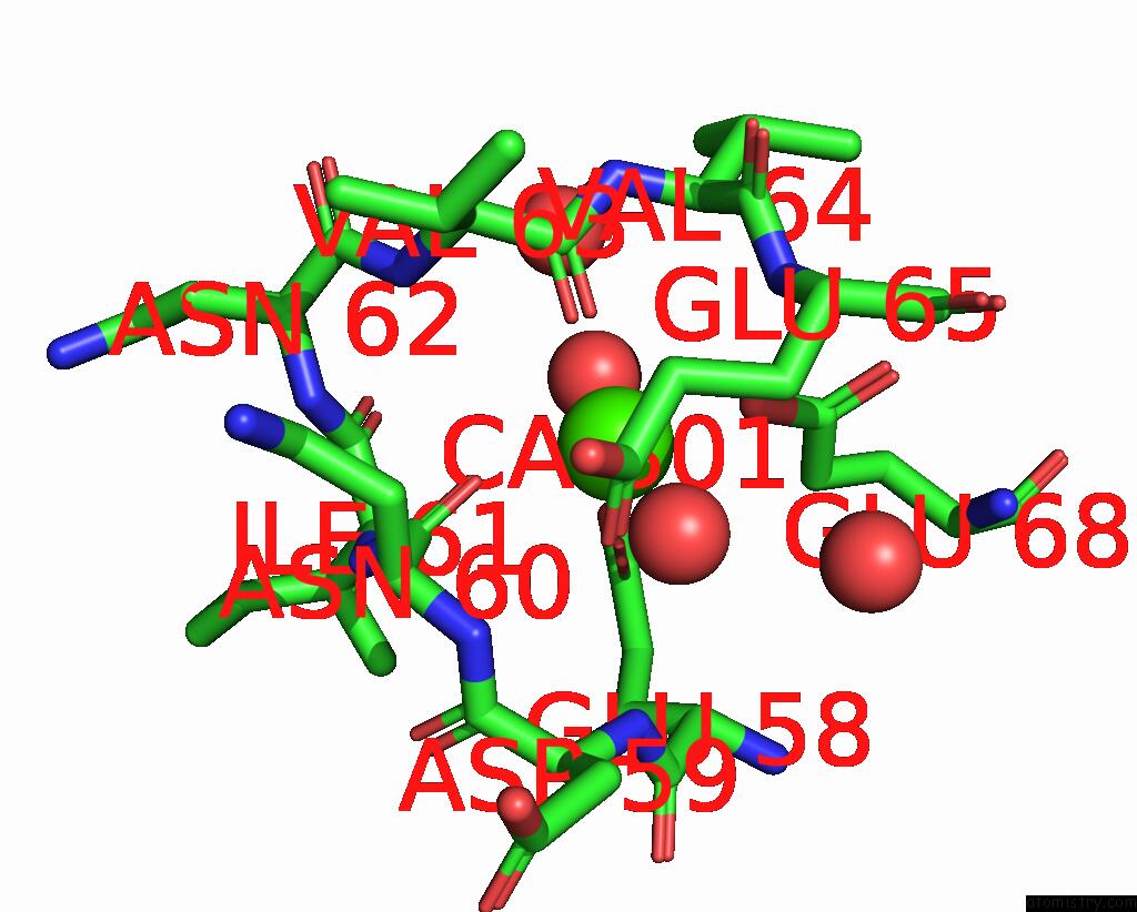
Mono view
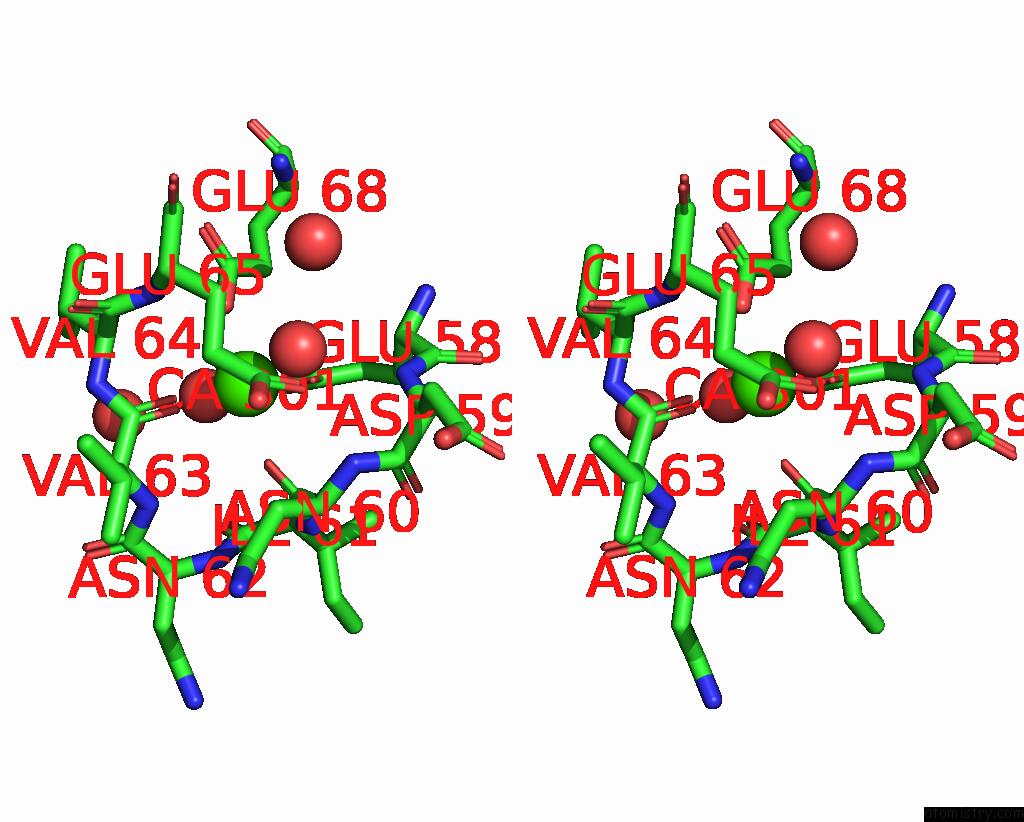
Stereo pair view
Mono view
Stereo pair view
A full contact list of Calcium with other atoms in the Ca binding
site number 1 of Anomalous Substructure of Trypsin (P3121) within 5.0Å range:
|
Reference:
C.Mueller-Dieckmann,
S.Panjikar,
A.Schmidt,
S.Mueller,
J.Kuper,
A.Geerlof,
M.Wilmanns,
R.K.Singh,
P.A.Tucker,
M.S.Weiss.
On the Routine Use of Soft X-Rays in Macromolecular Crystallography. Part IV. Efficient Determination of Anomalous Substructures in Biomacromolecules Using Longer X-Ray Wavelengths. Acta Crystallogr.,Sect.D V. 63 366 2007.
ISSN: ISSN 0907-4449
PubMed: 17327674
DOI: 10.1107/S0907444906055624
Page generated: Fri Jul 12 10:41:55 2024
ISSN: ISSN 0907-4449
PubMed: 17327674
DOI: 10.1107/S0907444906055624
Last articles
Zn in 9MJ5Zn in 9HNW
Zn in 9G0L
Zn in 9FNE
Zn in 9DZN
Zn in 9E0I
Zn in 9D32
Zn in 9DAK
Zn in 8ZXC
Zn in 8ZUF