Calcium »
PDB 2g8e-2gsm »
2gqt »
Calcium in PDB 2gqt: Crystal Structure of Udp-N-Acetylenolpyruvylglucosamine Reductase (Murb) From Thermus Caldophilus
Enzymatic activity of Crystal Structure of Udp-N-Acetylenolpyruvylglucosamine Reductase (Murb) From Thermus Caldophilus
All present enzymatic activity of Crystal Structure of Udp-N-Acetylenolpyruvylglucosamine Reductase (Murb) From Thermus Caldophilus:
1.1.1.158;
1.1.1.158;
Protein crystallography data
The structure of Crystal Structure of Udp-N-Acetylenolpyruvylglucosamine Reductase (Murb) From Thermus Caldophilus, PDB code: 2gqt
was solved by
M.-K.Kim,
S.H.Eom,
with X-Ray Crystallography technique. A brief refinement statistics is given in the table below:
| Resolution Low / High (Å) | 23.07 / 1.30 |
| Space group | P 1 |
| Cell size a, b, c (Å), α, β, γ (°) | 29.717, 46.501, 48.714, 64.83, 77.04, 84.40 |
| R / Rfree (%) | 18.7 / 20.9 |
Calcium Binding Sites:
The binding sites of Calcium atom in the Crystal Structure of Udp-N-Acetylenolpyruvylglucosamine Reductase (Murb) From Thermus Caldophilus
(pdb code 2gqt). This binding sites where shown within
5.0 Angstroms radius around Calcium atom.
In total only one binding site of Calcium was determined in the Crystal Structure of Udp-N-Acetylenolpyruvylglucosamine Reductase (Murb) From Thermus Caldophilus, PDB code: 2gqt:
In total only one binding site of Calcium was determined in the Crystal Structure of Udp-N-Acetylenolpyruvylglucosamine Reductase (Murb) From Thermus Caldophilus, PDB code: 2gqt:
Calcium binding site 1 out of 1 in 2gqt
Go back to
Calcium binding site 1 out
of 1 in the Crystal Structure of Udp-N-Acetylenolpyruvylglucosamine Reductase (Murb) From Thermus Caldophilus
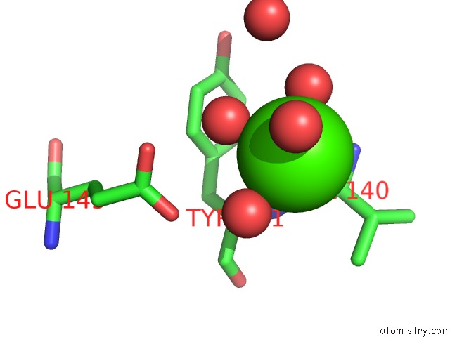
Mono view
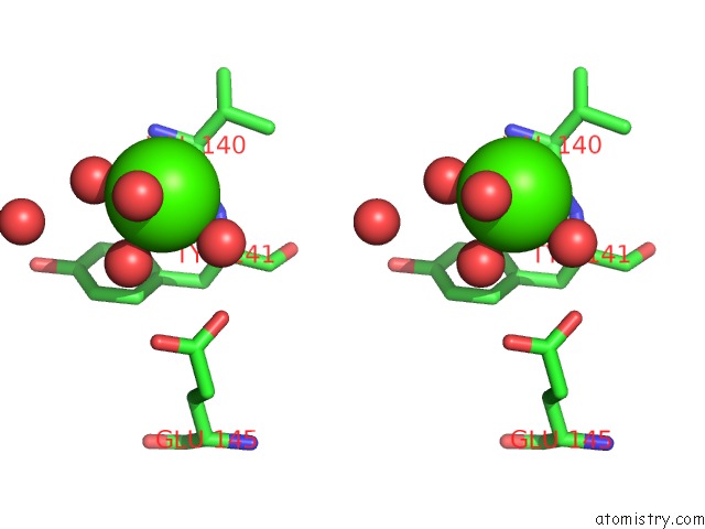
Stereo pair view
Mono view
Stereo pair view
A full contact list of Calcium with other atoms in the Ca binding
site number 1 of Crystal Structure of Udp-N-Acetylenolpyruvylglucosamine Reductase (Murb) From Thermus Caldophilus within 5.0Å range:
|
Reference:
M.-K.Kim,
M.K.Cho,
H.-E.Song,
D.Kim,
B.-H.Park,
J.H.Lee,
G.B.Kang,
S.H.Kim,
Y.J.Im,
D.-S.Lee,
S.H.Eom.
Crystal Structure of Udp-N-Acetylenolpyruvylglucosamine Reductase (Murb) From Thermus Caldophilus Proteins V. 66 751 2006.
ISSN: ISSN 0887-3585
PubMed: 17120230
DOI: 10.1002/PROT.21174
Page generated: Fri Jul 12 10:52:51 2024
ISSN: ISSN 0887-3585
PubMed: 17120230
DOI: 10.1002/PROT.21174
Last articles
Zn in 9J0NZn in 9J0O
Zn in 9J0P
Zn in 9FJX
Zn in 9EKB
Zn in 9C0F
Zn in 9CAH
Zn in 9CH0
Zn in 9CH3
Zn in 9CH1