Calcium »
PDB 2g8e-2gsm »
2gsm »
Calcium in PDB 2gsm: Catalytic Core (Subunits I and II) of Cytochrome C Oxidase From Rhodobacter Sphaeroides
Enzymatic activity of Catalytic Core (Subunits I and II) of Cytochrome C Oxidase From Rhodobacter Sphaeroides
All present enzymatic activity of Catalytic Core (Subunits I and II) of Cytochrome C Oxidase From Rhodobacter Sphaeroides:
1.9.3.1;
1.9.3.1;
Protein crystallography data
The structure of Catalytic Core (Subunits I and II) of Cytochrome C Oxidase From Rhodobacter Sphaeroides, PDB code: 2gsm
was solved by
L.Qin,
C.Hiser,
A.Mulichak,
R.M.Garavito,
S.Ferguson-Miller,
with X-Ray Crystallography technique. A brief refinement statistics is given in the table below:
| Resolution Low / High (Å) | 20.00 / 2.00 |
| Space group | P 21 21 21 |
| Cell size a, b, c (Å), α, β, γ (°) | 125.020, 131.639, 176.802, 90.00, 90.00, 90.00 |
| R / Rfree (%) | 21.4 / 23.2 |
Other elements in 2gsm:
The structure of Catalytic Core (Subunits I and II) of Cytochrome C Oxidase From Rhodobacter Sphaeroides also contains other interesting chemical elements:
| Magnesium | (Mg) | 2 atoms |
| Cadmium | (Cd) | 4 atoms |
| Iron | (Fe) | 4 atoms |
| Copper | (Cu) | 6 atoms |
Calcium Binding Sites:
The binding sites of Calcium atom in the Catalytic Core (Subunits I and II) of Cytochrome C Oxidase From Rhodobacter Sphaeroides
(pdb code 2gsm). This binding sites where shown within
5.0 Angstroms radius around Calcium atom.
In total 2 binding sites of Calcium where determined in the Catalytic Core (Subunits I and II) of Cytochrome C Oxidase From Rhodobacter Sphaeroides, PDB code: 2gsm:
Jump to Calcium binding site number: 1; 2;
In total 2 binding sites of Calcium where determined in the Catalytic Core (Subunits I and II) of Cytochrome C Oxidase From Rhodobacter Sphaeroides, PDB code: 2gsm:
Jump to Calcium binding site number: 1; 2;
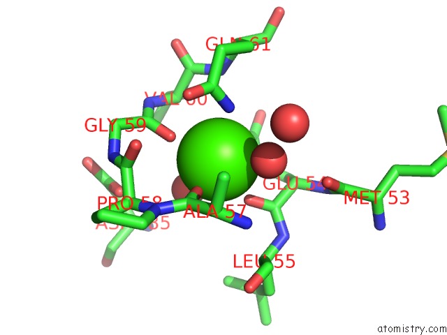
Calcium binding site 1 out of 2 in 2gsm
Go back to
Calcium binding site 1 out
of 2 in the Catalytic Core (Subunits I and II) of Cytochrome C Oxidase From Rhodobacter Sphaeroides
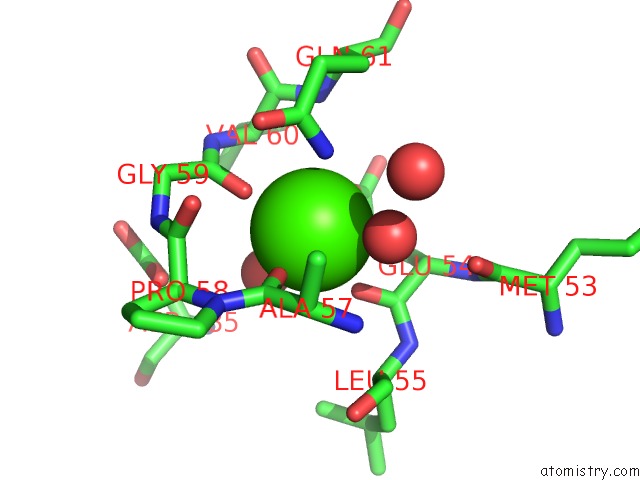
Mono view
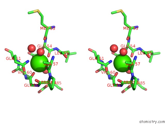
Stereo pair view
Mono view
Stereo pair view
A full contact list of Calcium with other atoms in the Ca binding
site number 1 of Catalytic Core (Subunits I and II) of Cytochrome C Oxidase From Rhodobacter Sphaeroides within 5.0Å range:
|
Calcium binding site 2 out of 2 in 2gsm
Go back to
Calcium binding site 2 out
of 2 in the Catalytic Core (Subunits I and II) of Cytochrome C Oxidase From Rhodobacter Sphaeroides
Mono view

Stereo pair view
Mono view
Stereo pair view
A full contact list of Calcium with other atoms in the Ca binding
site number 2 of Catalytic Core (Subunits I and II) of Cytochrome C Oxidase From Rhodobacter Sphaeroides within 5.0Å range:
|
Reference:
L.Qin,
C.Hiser,
A.Mulichak,
R.M.Garavito,
S.Ferguson-Miller.
Identification of Conserved Lipid/Detergent-Binding Sites in A High-Resolution Structure of the Membrane Protein Cytochrome C Oxidase. Proc.Natl.Acad.Sci.Usa V. 103 16117 2006.
ISSN: ISSN 0027-8424
PubMed: 17050688
DOI: 10.1073/PNAS.0606149103
Page generated: Tue Jul 8 05:50:21 2025
ISSN: ISSN 0027-8424
PubMed: 17050688
DOI: 10.1073/PNAS.0606149103
Last articles
Cl in 5JPVCl in 5JQ1
Cl in 5JOQ
Cl in 5JPD
Cl in 5JOO
Cl in 5JOH
Cl in 5JMR
Cl in 5JO3
Cl in 5JN0
Cl in 5JNY