Calcium »
PDB 2gsp-2hib »
2gwr »
Calcium in PDB 2gwr: Crystal Structure of the Response Regulator Protein Mtra From Mycobacterium Tuberculosis
Protein crystallography data
The structure of Crystal Structure of the Response Regulator Protein Mtra From Mycobacterium Tuberculosis, PDB code: 2gwr
was solved by
N.Friedland,
T.R.Mack,
M.Yu,
E.H.Bursey,
L.W.Hung,
A.M.Stock,
G.S.Waldo,
T.C.Terwilliger,
with X-Ray Crystallography technique. A brief refinement statistics is given in the table below:
| Resolution Low / High (Å) | 43.31 / 2.10 |
| Space group | P 21 21 21 |
| Cell size a, b, c (Å), α, β, γ (°) | 38.916, 56.598, 135.069, 90.00, 90.00, 90.00 |
| R / Rfree (%) | 20.4 / 24.4 |
Calcium Binding Sites:
The binding sites of Calcium atom in the Crystal Structure of the Response Regulator Protein Mtra From Mycobacterium Tuberculosis
(pdb code 2gwr). This binding sites where shown within
5.0 Angstroms radius around Calcium atom.
In total only one binding site of Calcium was determined in the Crystal Structure of the Response Regulator Protein Mtra From Mycobacterium Tuberculosis, PDB code: 2gwr:
In total only one binding site of Calcium was determined in the Crystal Structure of the Response Regulator Protein Mtra From Mycobacterium Tuberculosis, PDB code: 2gwr:
Calcium binding site 1 out of 1 in 2gwr
Go back to
Calcium binding site 1 out
of 1 in the Crystal Structure of the Response Regulator Protein Mtra From Mycobacterium Tuberculosis
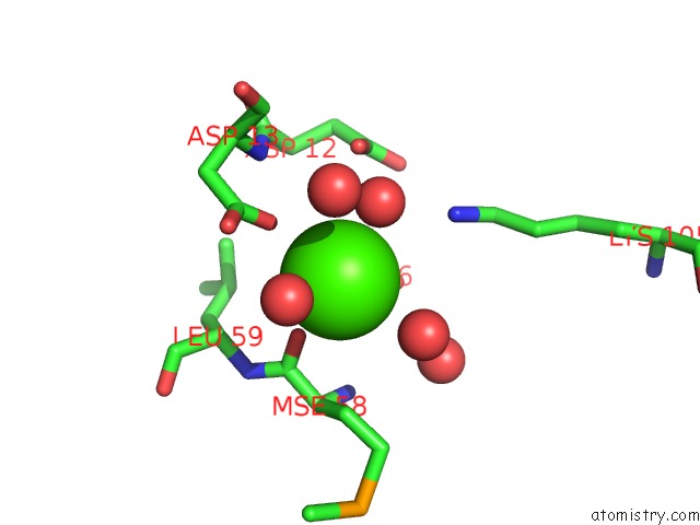
Mono view
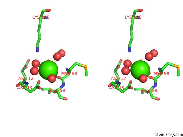
Stereo pair view
Mono view
Stereo pair view
A full contact list of Calcium with other atoms in the Ca binding
site number 1 of Crystal Structure of the Response Regulator Protein Mtra From Mycobacterium Tuberculosis within 5.0Å range:
|
Reference:
N.Friedland,
T.R.Mack,
M.Yu,
L.W.Hung,
T.C.Terwilliger,
G.S.Waldo,
A.M.Stock.
Domain Orientation in the Inactive Response Regulator Mycobacterium Tuberculosis Mtra Provides A Barrier to Activation. Biochemistry V. 46 6733 2007.
ISSN: ISSN 0006-2960
PubMed: 17511470
DOI: 10.1021/BI602546Q
Page generated: Fri Jul 12 10:57:03 2024
ISSN: ISSN 0006-2960
PubMed: 17511470
DOI: 10.1021/BI602546Q
Last articles
Zn in 9J0NZn in 9J0O
Zn in 9J0P
Zn in 9FJX
Zn in 9EKB
Zn in 9C0F
Zn in 9CAH
Zn in 9CH0
Zn in 9CH3
Zn in 9CH1