Calcium »
PDB 2gsp-2hib »
2h0k »
Calcium in PDB 2h0k: Crystal Structure of A Mutant of Rat Annexin A5
Protein crystallography data
The structure of Crystal Structure of A Mutant of Rat Annexin A5, PDB code: 2h0k
was solved by
T.Granier,
B.Langlois D'estaintot,
B.Gallois,
B.Tessier,
A.Brisson,
with X-Ray Crystallography technique. A brief refinement statistics is given in the table below:
| Resolution Low / High (Å) | 33.59 / 2.76 |
| Space group | P 1 21 1 |
| Cell size a, b, c (Å), α, β, γ (°) | 51.108, 67.184, 112.313, 90.00, 94.79, 90.00 |
| R / Rfree (%) | 19.7 / 26.1 |
Calcium Binding Sites:
Pages:
>>> Page 1 <<< Page 2, Binding sites: 11 - 20; Page 3, Binding sites: 21 - 22;Binding sites:
The binding sites of Calcium atom in the Crystal Structure of A Mutant of Rat Annexin A5 (pdb code 2h0k). This binding sites where shown within 5.0 Angstroms radius around Calcium atom.In total 22 binding sites of Calcium where determined in the Crystal Structure of A Mutant of Rat Annexin A5, PDB code: 2h0k:
Jump to Calcium binding site number: 1; 2; 3; 4; 5; 6; 7; 8; 9; 10;
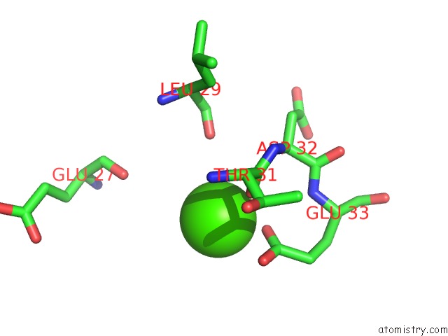
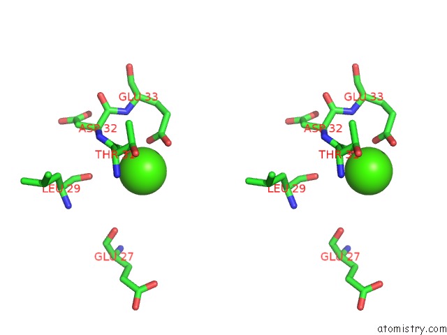
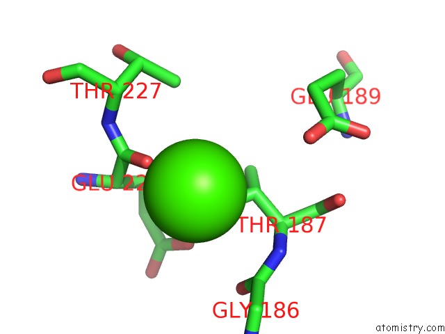
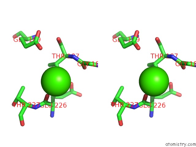
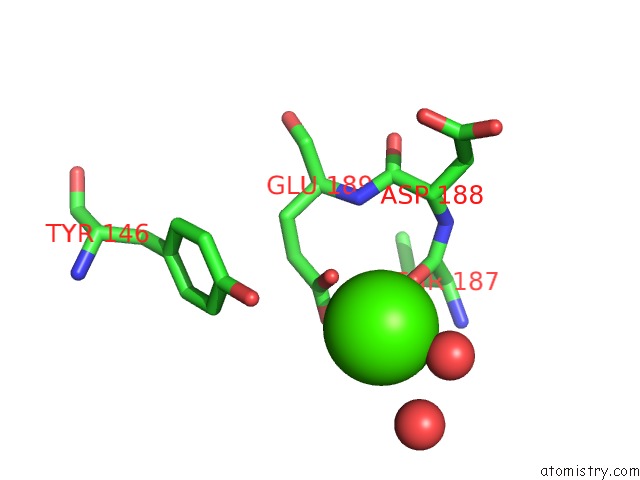
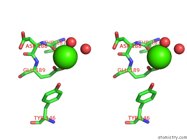
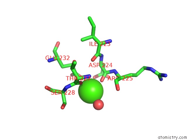
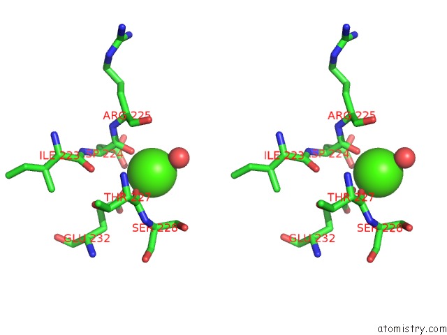
Calcium binding site 1 out of 22 in 2h0k
Go back to
Calcium binding site 1 out
of 22 in the Crystal Structure of A Mutant of Rat Annexin A5
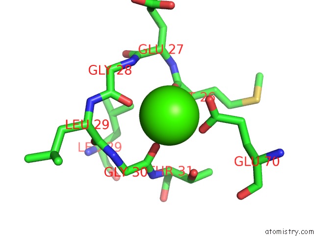
Mono view
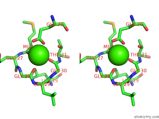
Stereo pair view
Mono view
Stereo pair view
A full contact list of Calcium with other atoms in the Ca binding
site number 1 of Crystal Structure of A Mutant of Rat Annexin A5 within 5.0Å range:
|
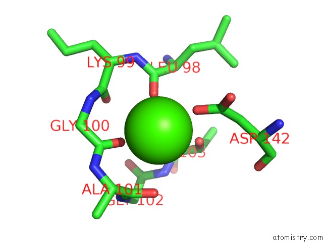
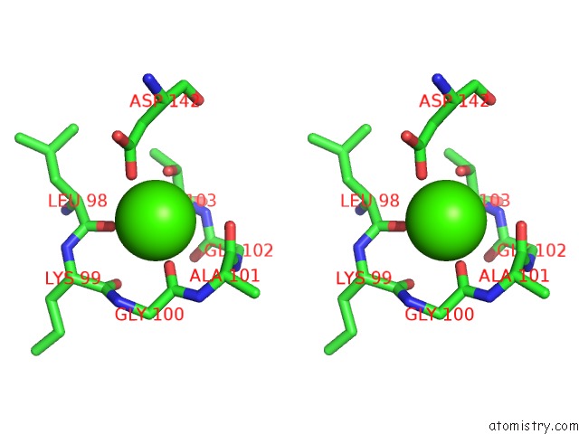
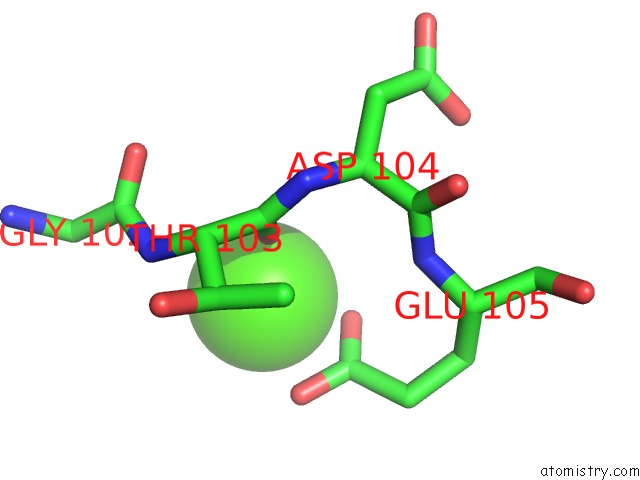
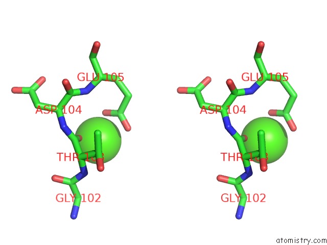
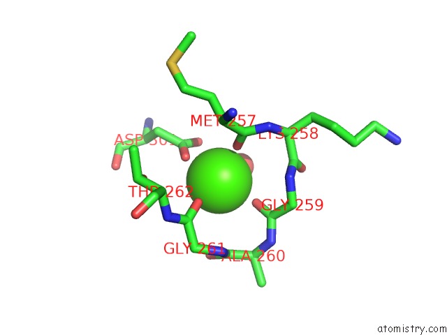
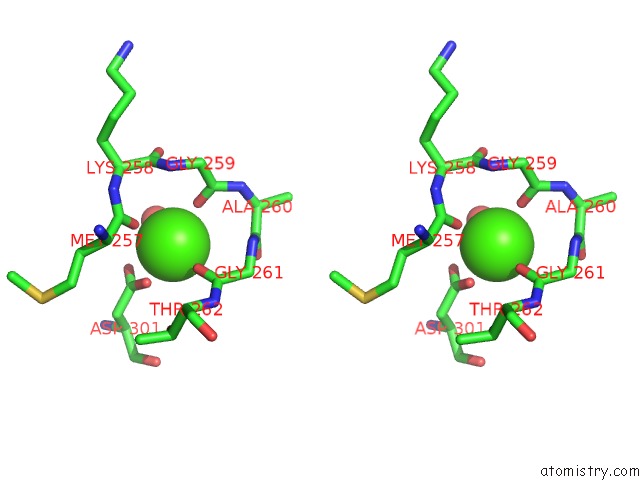
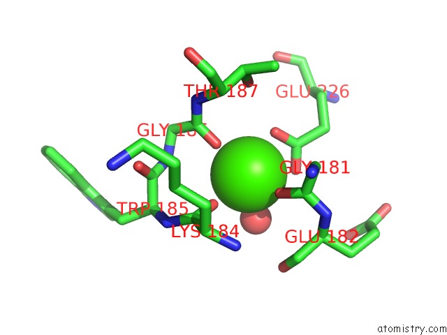
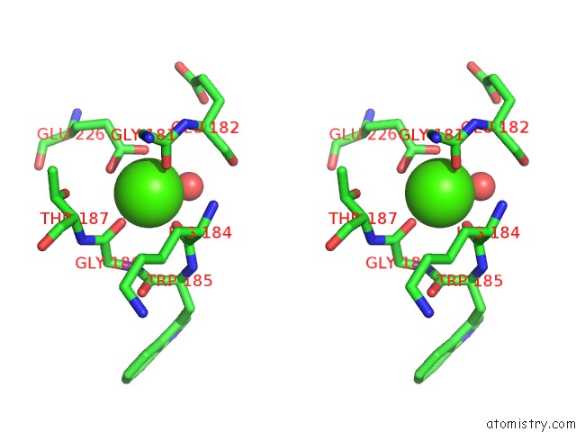
Calcium binding site 2 out of 22 in 2h0k
Go back to
Calcium binding site 2 out
of 22 in the Crystal Structure of A Mutant of Rat Annexin A5
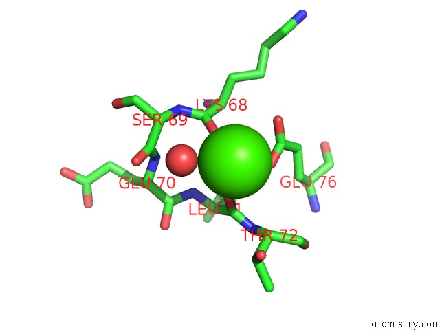
Mono view
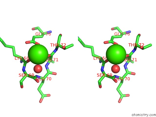
Stereo pair view
Mono view
Stereo pair view
A full contact list of Calcium with other atoms in the Ca binding
site number 2 of Crystal Structure of A Mutant of Rat Annexin A5 within 5.0Å range:
|
Calcium binding site 3 out of 22 in 2h0k
Go back to
Calcium binding site 3 out
of 22 in the Crystal Structure of A Mutant of Rat Annexin A5
Mono view
Stereo pair view
Mono view
Stereo pair view
A full contact list of Calcium with other atoms in the Ca binding
site number 3 of Crystal Structure of A Mutant of Rat Annexin A5 within 5.0Å range:
|
Calcium binding site 4 out of 22 in 2h0k
Go back to
Calcium binding site 4 out
of 22 in the Crystal Structure of A Mutant of Rat Annexin A5
Mono view
Stereo pair view
Mono view
Stereo pair view
A full contact list of Calcium with other atoms in the Ca binding
site number 4 of Crystal Structure of A Mutant of Rat Annexin A5 within 5.0Å range:
|
Calcium binding site 5 out of 22 in 2h0k
Go back to
Calcium binding site 5 out
of 22 in the Crystal Structure of A Mutant of Rat Annexin A5
Mono view
Stereo pair view
Mono view
Stereo pair view
A full contact list of Calcium with other atoms in the Ca binding
site number 5 of Crystal Structure of A Mutant of Rat Annexin A5 within 5.0Å range:
|
Calcium binding site 6 out of 22 in 2h0k
Go back to
Calcium binding site 6 out
of 22 in the Crystal Structure of A Mutant of Rat Annexin A5
Mono view
Stereo pair view
Mono view
Stereo pair view
A full contact list of Calcium with other atoms in the Ca binding
site number 6 of Crystal Structure of A Mutant of Rat Annexin A5 within 5.0Å range:
|
Calcium binding site 7 out of 22 in 2h0k
Go back to
Calcium binding site 7 out
of 22 in the Crystal Structure of A Mutant of Rat Annexin A5
Mono view
Stereo pair view
Mono view
Stereo pair view
A full contact list of Calcium with other atoms in the Ca binding
site number 7 of Crystal Structure of A Mutant of Rat Annexin A5 within 5.0Å range:
|
Calcium binding site 8 out of 22 in 2h0k
Go back to
Calcium binding site 8 out
of 22 in the Crystal Structure of A Mutant of Rat Annexin A5
Mono view
Stereo pair view
Mono view
Stereo pair view
A full contact list of Calcium with other atoms in the Ca binding
site number 8 of Crystal Structure of A Mutant of Rat Annexin A5 within 5.0Å range:
|
Calcium binding site 9 out of 22 in 2h0k
Go back to
Calcium binding site 9 out
of 22 in the Crystal Structure of A Mutant of Rat Annexin A5
Mono view
Stereo pair view
Mono view
Stereo pair view
A full contact list of Calcium with other atoms in the Ca binding
site number 9 of Crystal Structure of A Mutant of Rat Annexin A5 within 5.0Å range:
|
Calcium binding site 10 out of 22 in 2h0k
Go back to
Calcium binding site 10 out
of 22 in the Crystal Structure of A Mutant of Rat Annexin A5
Mono view
Stereo pair view
Mono view
Stereo pair view
A full contact list of Calcium with other atoms in the Ca binding
site number 10 of Crystal Structure of A Mutant of Rat Annexin A5 within 5.0Å range:
|
Reference:
A.Bouter,
C.Gounou,
R.Berat,
S.Tan,
B.Gallois,
T.Granier,
B.L.D'estaintot,
E.Poschl,
B.Brachvogel,
A.R.Brisson.
Annexin-A5 Assembled Into Two-Dimensional Arrays Promotes Cell Membrane Repair. Nat Commun V. 2 270 2011.
ISSN: ESSN 2041-1723
PubMed: 21468022
DOI: 10.1038/NCOMMS1270
Page generated: Fri Jul 12 10:57:34 2024
ISSN: ESSN 2041-1723
PubMed: 21468022
DOI: 10.1038/NCOMMS1270
Last articles
Zn in 9JYWZn in 9IR4
Zn in 9IR3
Zn in 9GMX
Zn in 9GMW
Zn in 9JEJ
Zn in 9ERF
Zn in 9ERE
Zn in 9EGV
Zn in 9EGW