Calcium »
PDB 2gsp-2hib »
2h0l »
Calcium in PDB 2h0l: Crystal Structure of A Mutant of Rat Annexin A5
Protein crystallography data
The structure of Crystal Structure of A Mutant of Rat Annexin A5, PDB code: 2h0l
was solved by
B.Langlois D'estaintot,
B.Gallois,
T.Granier,
B.Tessier,
A.Brisson,
with X-Ray Crystallography technique. A brief refinement statistics is given in the table below:
| Resolution Low / High (Å) | 32.76 / 2.59 |
| Space group | P 21 21 2 |
| Cell size a, b, c (Å), α, β, γ (°) | 77.918, 78.959, 60.458, 90.00, 90.00, 90.00 |
| R / Rfree (%) | 18.7 / 27.8 |
Calcium Binding Sites:
The binding sites of Calcium atom in the Crystal Structure of A Mutant of Rat Annexin A5
(pdb code 2h0l). This binding sites where shown within
5.0 Angstroms radius around Calcium atom.
In total 3 binding sites of Calcium where determined in the Crystal Structure of A Mutant of Rat Annexin A5, PDB code: 2h0l:
Jump to Calcium binding site number: 1; 2; 3;
In total 3 binding sites of Calcium where determined in the Crystal Structure of A Mutant of Rat Annexin A5, PDB code: 2h0l:
Jump to Calcium binding site number: 1; 2; 3;
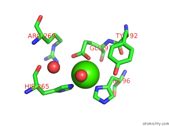
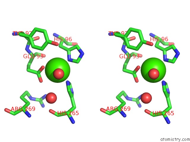
Calcium binding site 1 out of 3 in 2h0l
Go back to
Calcium binding site 1 out
of 3 in the Crystal Structure of A Mutant of Rat Annexin A5
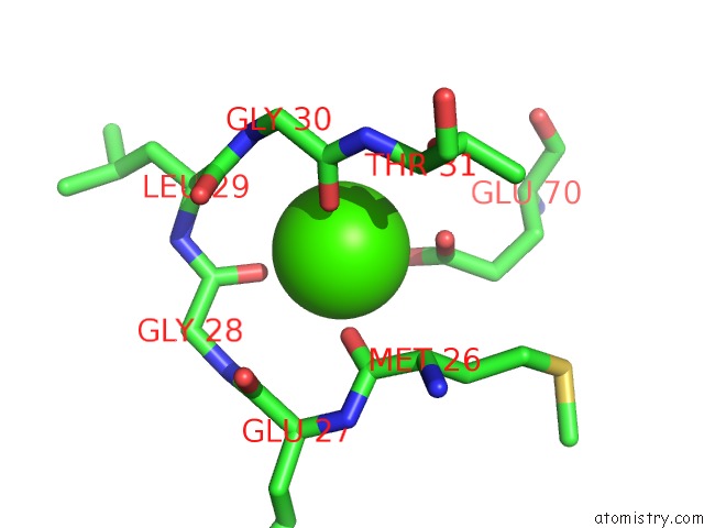
Mono view
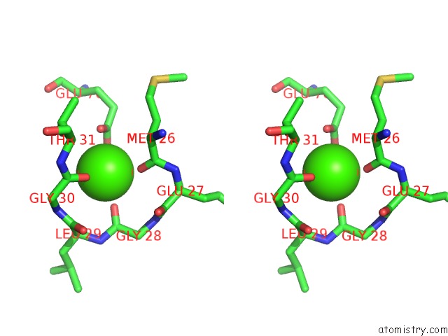
Stereo pair view
Mono view
Stereo pair view
A full contact list of Calcium with other atoms in the Ca binding
site number 1 of Crystal Structure of A Mutant of Rat Annexin A5 within 5.0Å range:
|
Calcium binding site 2 out of 3 in 2h0l
Go back to
Calcium binding site 2 out
of 3 in the Crystal Structure of A Mutant of Rat Annexin A5
Mono view
Stereo pair view
Mono view
Stereo pair view
A full contact list of Calcium with other atoms in the Ca binding
site number 2 of Crystal Structure of A Mutant of Rat Annexin A5 within 5.0Å range:
|
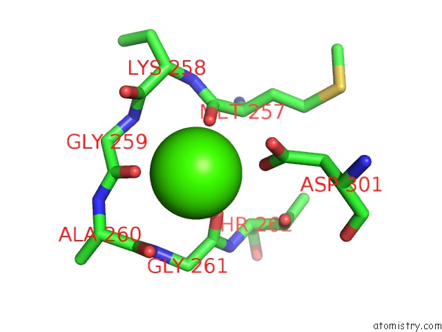
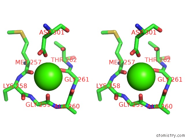
Calcium binding site 3 out of 3 in 2h0l
Go back to
Calcium binding site 3 out
of 3 in the Crystal Structure of A Mutant of Rat Annexin A5
Mono view
Stereo pair view
Mono view
Stereo pair view
A full contact list of Calcium with other atoms in the Ca binding
site number 3 of Crystal Structure of A Mutant of Rat Annexin A5 within 5.0Å range:
|
Reference:
A.Brisson,
T.Granier,
B.Langlois D'estaintot,
B.Gallois,
B.Tessier.
Identification of the Residues Involved in the Formation of Annexin V Trimers Within 2D and 3D Crystals To Be Published.
Page generated: Fri Jul 12 10:57:58 2024
Last articles
Zn in 9J0NZn in 9J0O
Zn in 9J0P
Zn in 9FJX
Zn in 9EKB
Zn in 9C0F
Zn in 9CAH
Zn in 9CH0
Zn in 9CH3
Zn in 9CH1