Calcium »
PDB 2gsp-2hib »
2h2t »
Calcium in PDB 2h2t: CD23 Lectin Domain, Calcium 2+-Bound
Protein crystallography data
The structure of CD23 Lectin Domain, Calcium 2+-Bound, PDB code: 2h2t
was solved by
B.A.Wurzburg,
with X-Ray Crystallography technique. A brief refinement statistics is given in the table below:
| Resolution Low / High (Å) | 38.63 / 1.30 |
| Space group | P 21 21 21 |
| Cell size a, b, c (Å), α, β, γ (°) | 40.310, 51.261, 58.768, 90.00, 90.00, 90.00 |
| R / Rfree (%) | 16.8 / 20 |
Calcium Binding Sites:
The binding sites of Calcium atom in the CD23 Lectin Domain, Calcium 2+-Bound
(pdb code 2h2t). This binding sites where shown within
5.0 Angstroms radius around Calcium atom.
In total only one binding site of Calcium was determined in the CD23 Lectin Domain, Calcium 2+-Bound, PDB code: 2h2t:
In total only one binding site of Calcium was determined in the CD23 Lectin Domain, Calcium 2+-Bound, PDB code: 2h2t:
Calcium binding site 1 out of 1 in 2h2t
Go back to
Calcium binding site 1 out
of 1 in the CD23 Lectin Domain, Calcium 2+-Bound
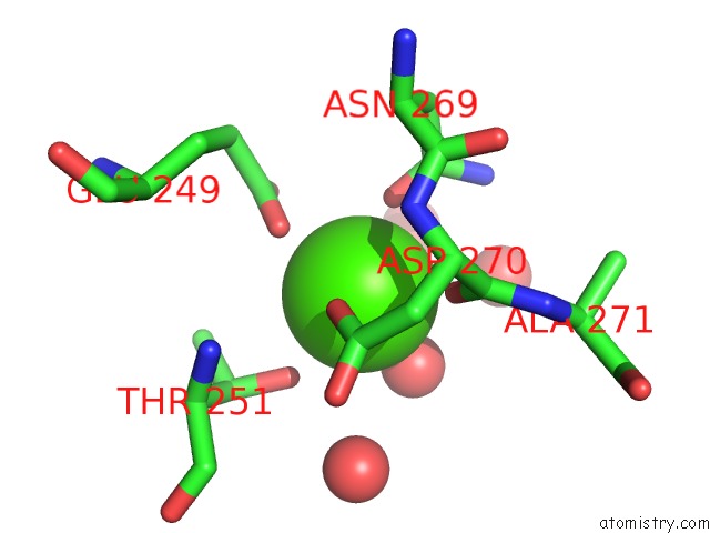
Mono view
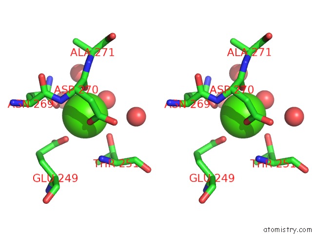
Stereo pair view
Mono view
Stereo pair view
A full contact list of Calcium with other atoms in the Ca binding
site number 1 of CD23 Lectin Domain, Calcium 2+-Bound within 5.0Å range:
|
Reference:
B.A.Wurzburg,
S.S.Tarchevskaya,
T.S.Jardetzky.
Structural Changes in the Lectin Domain of CD23, the Low-Affinity Ige Receptor, Upon Calcium Binding. Structure V. 14 1049 2006.
ISSN: ISSN 0969-2126
PubMed: 16765898
DOI: 10.1016/J.STR.2006.03.017
Page generated: Fri Jul 12 12:43:12 2024
ISSN: ISSN 0969-2126
PubMed: 16765898
DOI: 10.1016/J.STR.2006.03.017
Last articles
Zn in 9J0NZn in 9J0O
Zn in 9J0P
Zn in 9FJX
Zn in 9EKB
Zn in 9C0F
Zn in 9CAH
Zn in 9CH0
Zn in 9CH3
Zn in 9CH1