Calcium »
PDB 2gsp-2hib »
2h43 »
Calcium in PDB 2h43: Crystal Structure of Human Fragment D Complexed with Ala-His-Arg-Pro- Amide
Protein crystallography data
The structure of Crystal Structure of Human Fragment D Complexed with Ala-His-Arg-Pro- Amide, PDB code: 2h43
was solved by
R.F.Doolittle,
L.Pandi,
with X-Ray Crystallography technique. A brief refinement statistics is given in the table below:
| Resolution Low / High (Å) | 30.00 / 2.70 |
| Space group | P 1 21 1 |
| Cell size a, b, c (Å), α, β, γ (°) | 105.000, 47.900, 171.400, 90.00, 105.41, 90.00 |
| R / Rfree (%) | 25.1 / 30.7 |
Calcium Binding Sites:
The binding sites of Calcium atom in the Crystal Structure of Human Fragment D Complexed with Ala-His-Arg-Pro- Amide
(pdb code 2h43). This binding sites where shown within
5.0 Angstroms radius around Calcium atom.
In total 6 binding sites of Calcium where determined in the Crystal Structure of Human Fragment D Complexed with Ala-His-Arg-Pro- Amide, PDB code: 2h43:
Jump to Calcium binding site number: 1; 2; 3; 4; 5; 6;
In total 6 binding sites of Calcium where determined in the Crystal Structure of Human Fragment D Complexed with Ala-His-Arg-Pro- Amide, PDB code: 2h43:
Jump to Calcium binding site number: 1; 2; 3; 4; 5; 6;
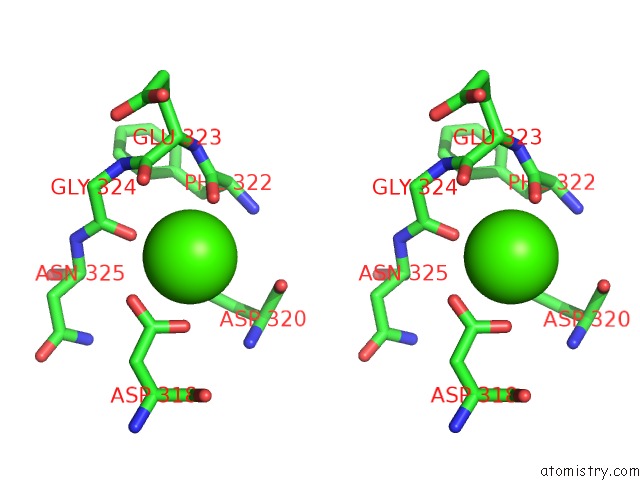
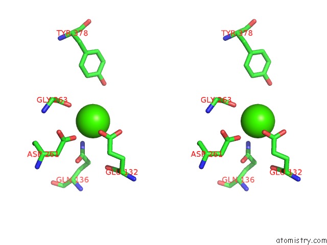
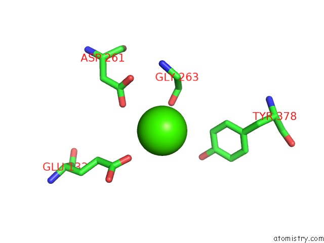
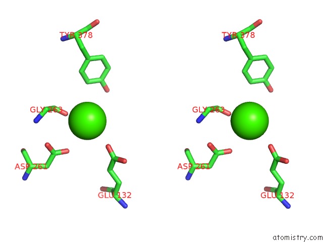
Calcium binding site 1 out of 6 in 2h43
Go back to
Calcium binding site 1 out
of 6 in the Crystal Structure of Human Fragment D Complexed with Ala-His-Arg-Pro- Amide

Mono view
Stereo pair view
Mono view
Stereo pair view
A full contact list of Calcium with other atoms in the Ca binding
site number 1 of Crystal Structure of Human Fragment D Complexed with Ala-His-Arg-Pro- Amide within 5.0Å range:
|
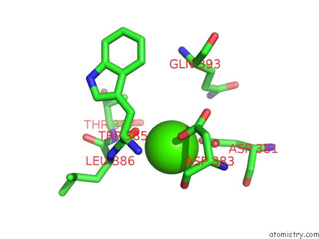
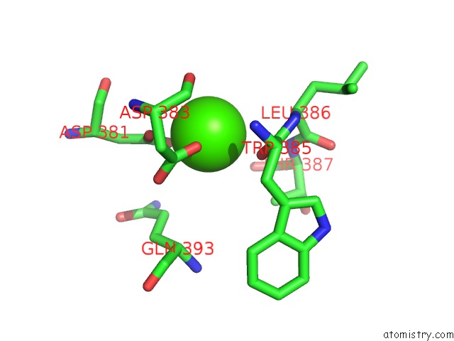
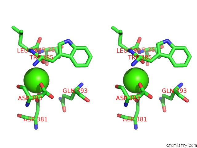
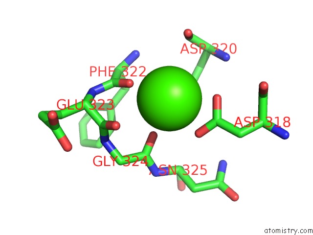
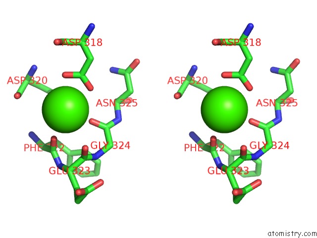
Calcium binding site 2 out of 6 in 2h43
Go back to
Calcium binding site 2 out
of 6 in the Crystal Structure of Human Fragment D Complexed with Ala-His-Arg-Pro- Amide
Mono view
Stereo pair view
Mono view
Stereo pair view
A full contact list of Calcium with other atoms in the Ca binding
site number 2 of Crystal Structure of Human Fragment D Complexed with Ala-His-Arg-Pro- Amide within 5.0Å range:
|
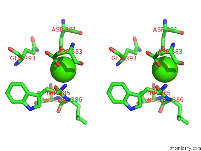
Calcium binding site 3 out of 6 in 2h43
Go back to
Calcium binding site 3 out
of 6 in the Crystal Structure of Human Fragment D Complexed with Ala-His-Arg-Pro- Amide
Mono view
Stereo pair view
Mono view
Stereo pair view
A full contact list of Calcium with other atoms in the Ca binding
site number 3 of Crystal Structure of Human Fragment D Complexed with Ala-His-Arg-Pro- Amide within 5.0Å range:
|
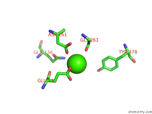
Calcium binding site 4 out of 6 in 2h43
Go back to
Calcium binding site 4 out
of 6 in the Crystal Structure of Human Fragment D Complexed with Ala-His-Arg-Pro- Amide
Mono view
Stereo pair view
Mono view
Stereo pair view
A full contact list of Calcium with other atoms in the Ca binding
site number 4 of Crystal Structure of Human Fragment D Complexed with Ala-His-Arg-Pro- Amide within 5.0Å range:
|
Calcium binding site 5 out of 6 in 2h43
Go back to
Calcium binding site 5 out
of 6 in the Crystal Structure of Human Fragment D Complexed with Ala-His-Arg-Pro- Amide
Mono view
Stereo pair view
Mono view
Stereo pair view
A full contact list of Calcium with other atoms in the Ca binding
site number 5 of Crystal Structure of Human Fragment D Complexed with Ala-His-Arg-Pro- Amide within 5.0Å range:
|
Calcium binding site 6 out of 6 in 2h43
Go back to
Calcium binding site 6 out
of 6 in the Crystal Structure of Human Fragment D Complexed with Ala-His-Arg-Pro- Amide
Mono view
Stereo pair view
Mono view
Stereo pair view
A full contact list of Calcium with other atoms in the Ca binding
site number 6 of Crystal Structure of Human Fragment D Complexed with Ala-His-Arg-Pro- Amide within 5.0Å range:
|
Reference:
R.F.Doolittle,
A.Chen,
L.Pandi.
Differences in Binding Specificity For the Homologous Gamma- and Beta-Chain "Holes" on Fibrinogen: Exclusive Binding of Ala-His-Arg-Pro-Amide By the Beta-Chain Hole. Biochemistry V. 45 13962 2006.
ISSN: ISSN 0006-2960
PubMed: 17115691
DOI: 10.1021/BI061219E
Page generated: Tue Jul 8 05:55:17 2025
ISSN: ISSN 0006-2960
PubMed: 17115691
DOI: 10.1021/BI061219E
Last articles
Cl in 5JS6Cl in 5JQG
Cl in 5JQS
Cl in 5JPI
Cl in 5JPH
Cl in 5JQ4
Cl in 5JQ7
Cl in 5JPV
Cl in 5JQ1
Cl in 5JOQ