Calcium »
PDB 2gsp-2hib »
2hcn »
Calcium in PDB 2hcn: Crystal Structure of Rna Dependent Rna Polymerase Domain From West Nile Virus
Enzymatic activity of Crystal Structure of Rna Dependent Rna Polymerase Domain From West Nile Virus
All present enzymatic activity of Crystal Structure of Rna Dependent Rna Polymerase Domain From West Nile Virus:
2.7.7.48;
2.7.7.48;
Protein crystallography data
The structure of Crystal Structure of Rna Dependent Rna Polymerase Domain From West Nile Virus, PDB code: 2hcn
was solved by
M.P.Egloff,
H.Malet,
Marseilles Structural Genomics Program @ Afmb(Msgp),
with X-Ray Crystallography technique. A brief refinement statistics is given in the table below:
| Resolution Low / High (Å) | 34.82 / 2.35 |
| Space group | P 43 |
| Cell size a, b, c (Å), α, β, γ (°) | 110.064, 110.064, 68.572, 90.00, 90.00, 90.00 |
| R / Rfree (%) | 20.7 / 25.9 |
Other elements in 2hcn:
The structure of Crystal Structure of Rna Dependent Rna Polymerase Domain From West Nile Virus also contains other interesting chemical elements:
| Zinc | (Zn) | 2 atoms |
Calcium Binding Sites:
The binding sites of Calcium atom in the Crystal Structure of Rna Dependent Rna Polymerase Domain From West Nile Virus
(pdb code 2hcn). This binding sites where shown within
5.0 Angstroms radius around Calcium atom.
In total only one binding site of Calcium was determined in the Crystal Structure of Rna Dependent Rna Polymerase Domain From West Nile Virus, PDB code: 2hcn:
In total only one binding site of Calcium was determined in the Crystal Structure of Rna Dependent Rna Polymerase Domain From West Nile Virus, PDB code: 2hcn:
Calcium binding site 1 out of 1 in 2hcn
Go back to
Calcium binding site 1 out
of 1 in the Crystal Structure of Rna Dependent Rna Polymerase Domain From West Nile Virus
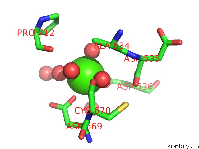
Mono view
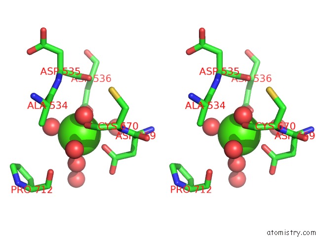
Stereo pair view
Mono view
Stereo pair view
A full contact list of Calcium with other atoms in the Ca binding
site number 1 of Crystal Structure of Rna Dependent Rna Polymerase Domain From West Nile Virus within 5.0Å range:
|
Reference:
H.Malet,
M.P.Egloff,
B.Selisko,
R.E.Butcher,
P.J.Wright,
M.Roberts,
A.Gruez,
G.Sulzenbacher,
C.Vonrhein,
G.Bricogne,
J.M.Mackenzie,
A.A.Khromykh,
A.D.Davidson,
B.Canard.
Crystal Structure of the Rna Polymerase Domain of the West Nile Virus Non-Structural Protein 5 J.Biol.Chem. V. 282 10678 2007.
ISSN: ISSN 0021-9258
PubMed: 17287213
DOI: 10.1074/JBC.M607273200
Page generated: Fri Jul 12 11:03:10 2024
ISSN: ISSN 0021-9258
PubMed: 17287213
DOI: 10.1074/JBC.M607273200
Last articles
Zn in 9MJ5Zn in 9HNW
Zn in 9G0L
Zn in 9FNE
Zn in 9DZN
Zn in 9E0I
Zn in 9D32
Zn in 9DAK
Zn in 8ZXC
Zn in 8ZUF