Calcium »
PDB 2hih-2i52 »
2hok »
Calcium in PDB 2hok: Crystal Structure of An E. Coli Thi-Box Riboswitch Bound to Thiamine Pyrophosphate, Calcium Ions
Protein crystallography data
The structure of Crystal Structure of An E. Coli Thi-Box Riboswitch Bound to Thiamine Pyrophosphate, Calcium Ions, PDB code: 2hok
was solved by
T.E.Edwards,
A.R.Ferre-D'amare,
with X-Ray Crystallography technique. A brief refinement statistics is given in the table below:
| Resolution Low / High (Å) | 30.00 / 3.20 |
| Space group | P 32 1 2 |
| Cell size a, b, c (Å), α, β, γ (°) | 66.300, 66.300, 101.600, 90.00, 90.00, 120.00 |
| R / Rfree (%) | 21.4 / 29.1 |
Calcium Binding Sites:
The binding sites of Calcium atom in the Crystal Structure of An E. Coli Thi-Box Riboswitch Bound to Thiamine Pyrophosphate, Calcium Ions
(pdb code 2hok). This binding sites where shown within
5.0 Angstroms radius around Calcium atom.
In total 7 binding sites of Calcium where determined in the Crystal Structure of An E. Coli Thi-Box Riboswitch Bound to Thiamine Pyrophosphate, Calcium Ions, PDB code: 2hok:
Jump to Calcium binding site number: 1; 2; 3; 4; 5; 6; 7;
In total 7 binding sites of Calcium where determined in the Crystal Structure of An E. Coli Thi-Box Riboswitch Bound to Thiamine Pyrophosphate, Calcium Ions, PDB code: 2hok:
Jump to Calcium binding site number: 1; 2; 3; 4; 5; 6; 7;
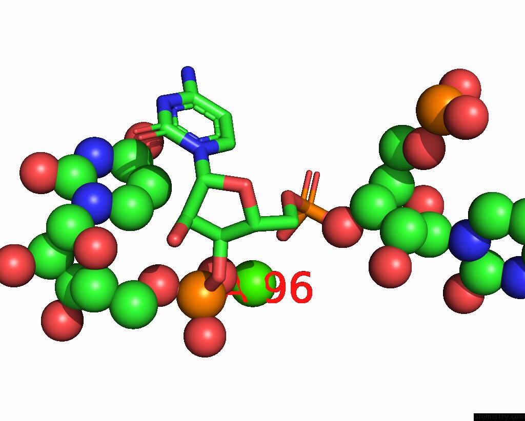
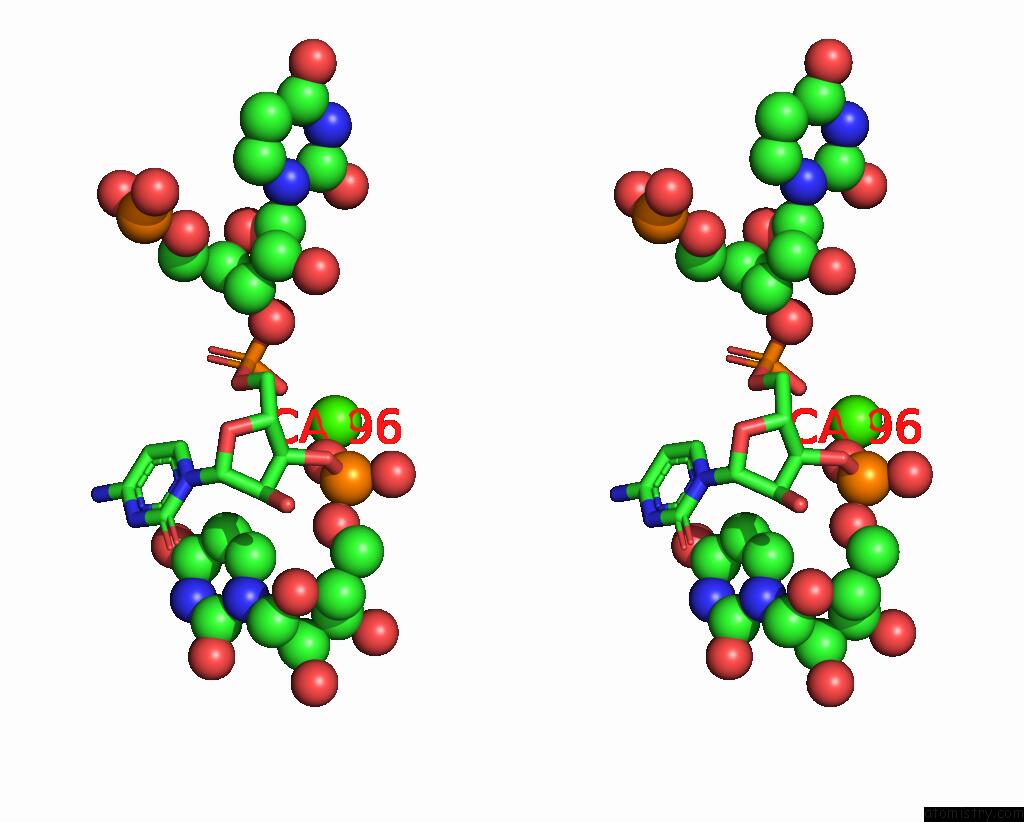
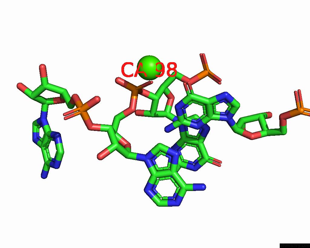
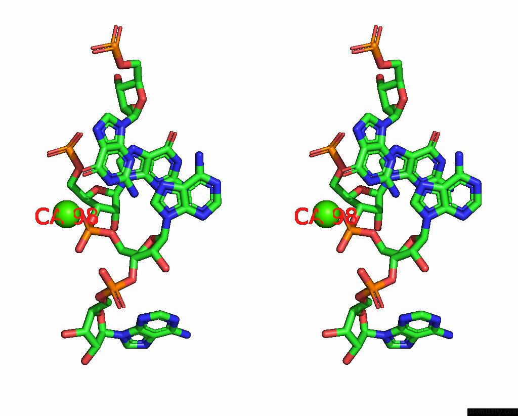
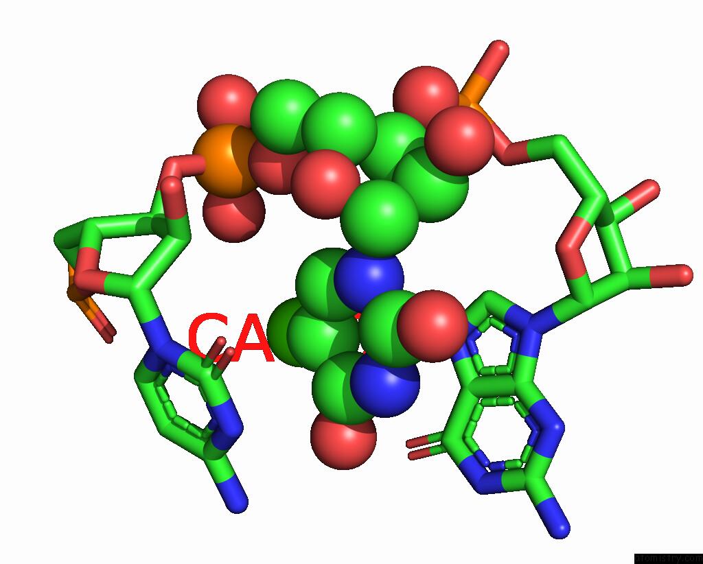
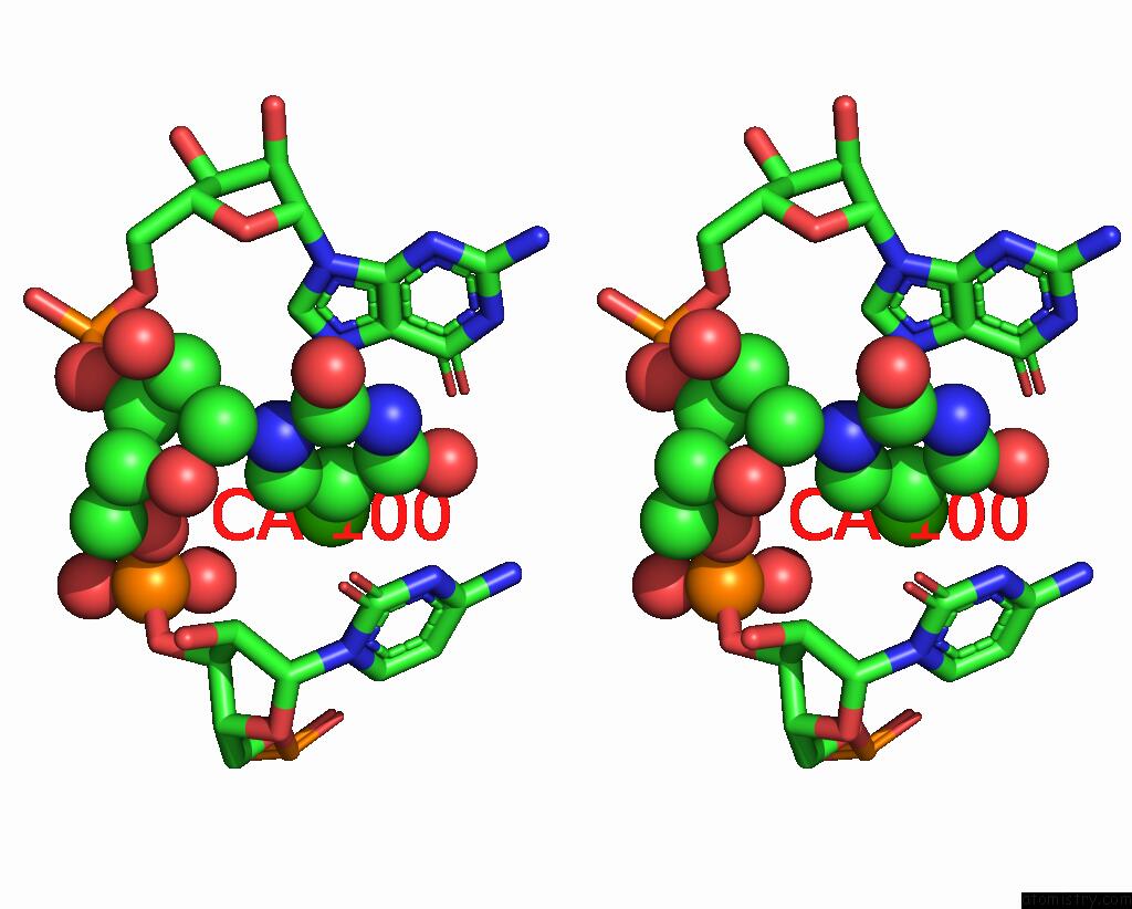
Calcium binding site 1 out of 7 in 2hok
Go back to
Calcium binding site 1 out
of 7 in the Crystal Structure of An E. Coli Thi-Box Riboswitch Bound to Thiamine Pyrophosphate, Calcium Ions
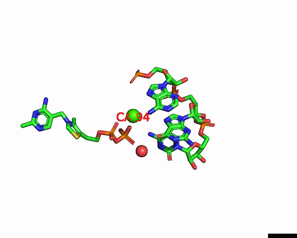
Mono view
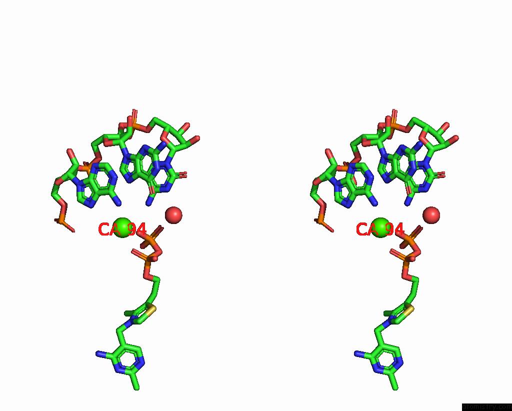
Stereo pair view
Mono view
Stereo pair view
A full contact list of Calcium with other atoms in the Ca binding
site number 1 of Crystal Structure of An E. Coli Thi-Box Riboswitch Bound to Thiamine Pyrophosphate, Calcium Ions within 5.0Å range:
|
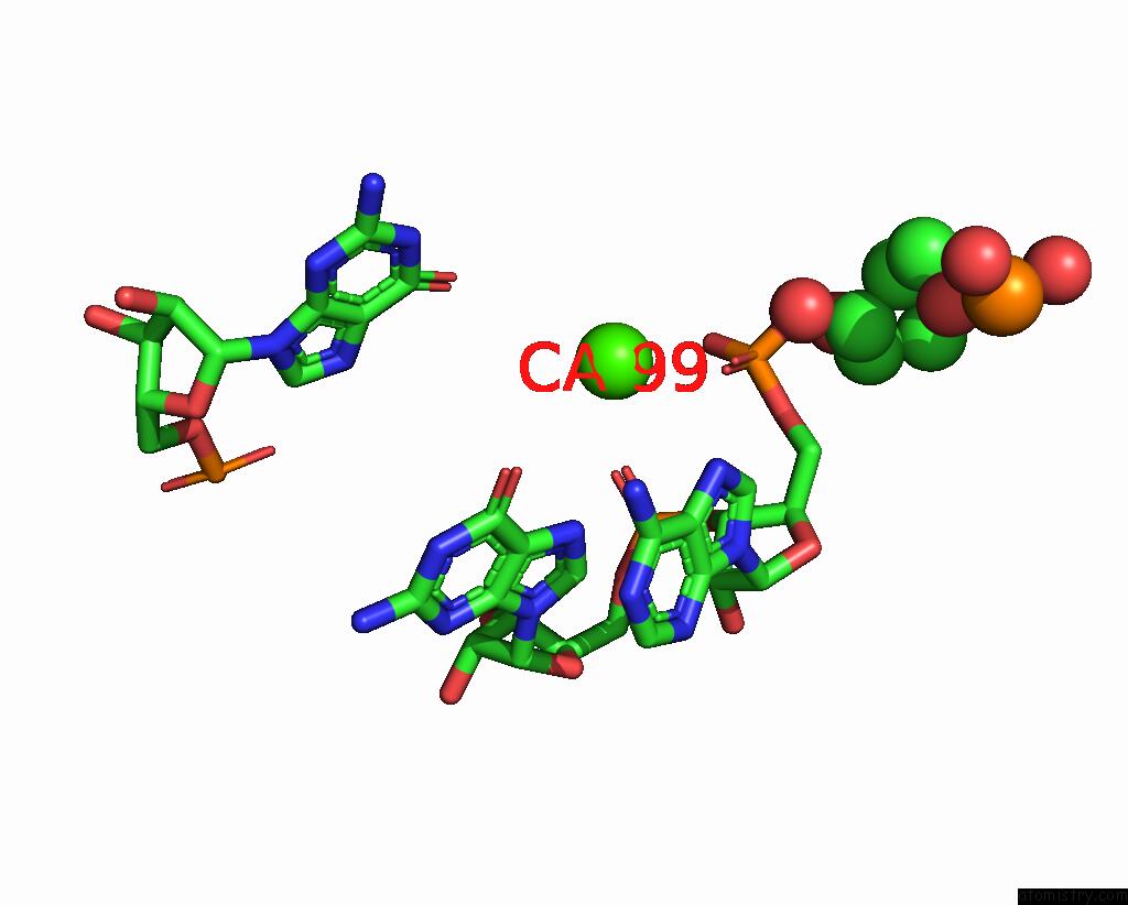
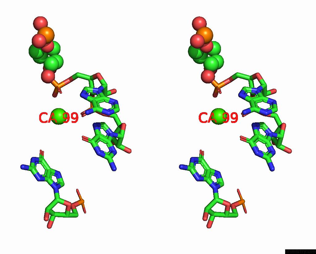
Calcium binding site 2 out of 7 in 2hok
Go back to
Calcium binding site 2 out
of 7 in the Crystal Structure of An E. Coli Thi-Box Riboswitch Bound to Thiamine Pyrophosphate, Calcium Ions
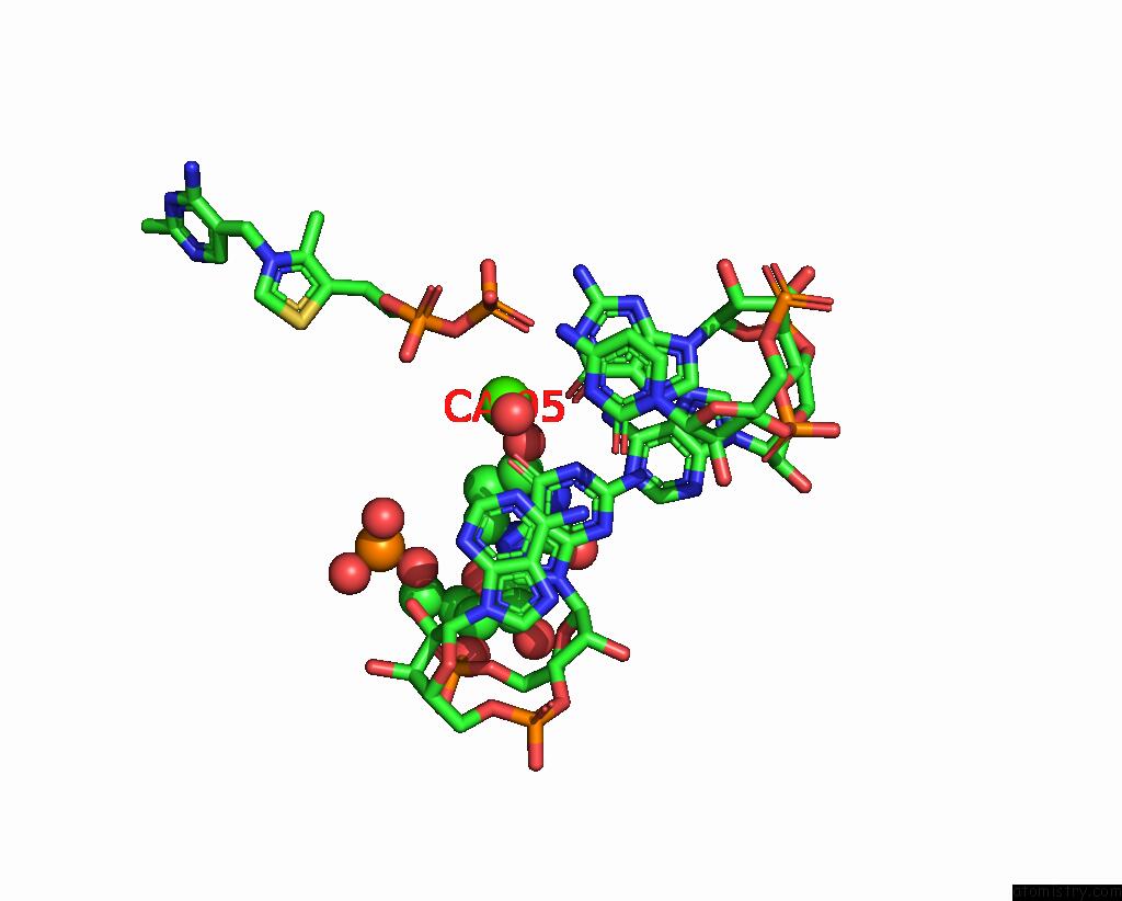
Mono view
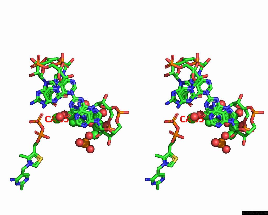
Stereo pair view
Mono view
Stereo pair view
A full contact list of Calcium with other atoms in the Ca binding
site number 2 of Crystal Structure of An E. Coli Thi-Box Riboswitch Bound to Thiamine Pyrophosphate, Calcium Ions within 5.0Å range:
|
Calcium binding site 3 out of 7 in 2hok
Go back to
Calcium binding site 3 out
of 7 in the Crystal Structure of An E. Coli Thi-Box Riboswitch Bound to Thiamine Pyrophosphate, Calcium Ions
Mono view
Stereo pair view
Mono view
Stereo pair view
A full contact list of Calcium with other atoms in the Ca binding
site number 3 of Crystal Structure of An E. Coli Thi-Box Riboswitch Bound to Thiamine Pyrophosphate, Calcium Ions within 5.0Å range:
|
Calcium binding site 4 out of 7 in 2hok
Go back to
Calcium binding site 4 out
of 7 in the Crystal Structure of An E. Coli Thi-Box Riboswitch Bound to Thiamine Pyrophosphate, Calcium Ions
Mono view
Stereo pair view
Mono view
Stereo pair view
A full contact list of Calcium with other atoms in the Ca binding
site number 4 of Crystal Structure of An E. Coli Thi-Box Riboswitch Bound to Thiamine Pyrophosphate, Calcium Ions within 5.0Å range:
|
Calcium binding site 5 out of 7 in 2hok
Go back to
Calcium binding site 5 out
of 7 in the Crystal Structure of An E. Coli Thi-Box Riboswitch Bound to Thiamine Pyrophosphate, Calcium Ions
Mono view
Stereo pair view
Mono view
Stereo pair view
A full contact list of Calcium with other atoms in the Ca binding
site number 5 of Crystal Structure of An E. Coli Thi-Box Riboswitch Bound to Thiamine Pyrophosphate, Calcium Ions within 5.0Å range:
|
Calcium binding site 6 out of 7 in 2hok
Go back to
Calcium binding site 6 out
of 7 in the Crystal Structure of An E. Coli Thi-Box Riboswitch Bound to Thiamine Pyrophosphate, Calcium Ions
Mono view
Stereo pair view
Mono view
Stereo pair view
A full contact list of Calcium with other atoms in the Ca binding
site number 6 of Crystal Structure of An E. Coli Thi-Box Riboswitch Bound to Thiamine Pyrophosphate, Calcium Ions within 5.0Å range:
|
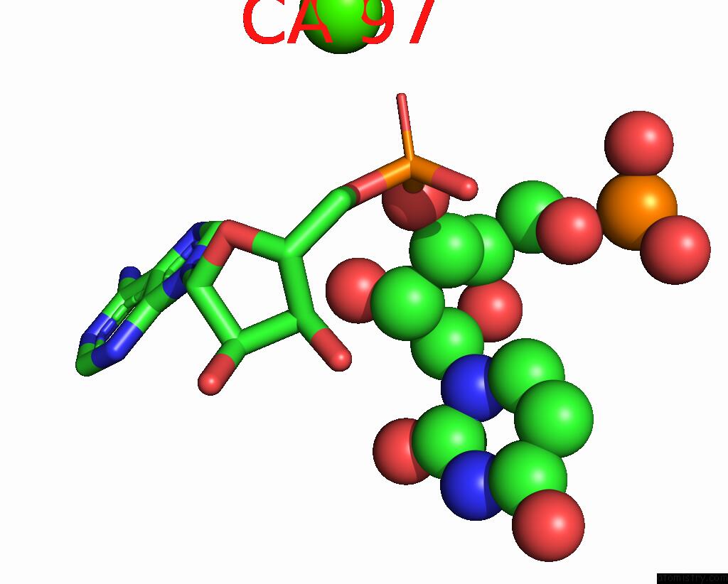
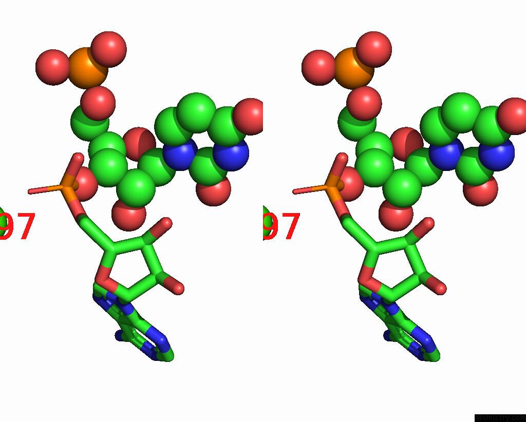
Calcium binding site 7 out of 7 in 2hok
Go back to
Calcium binding site 7 out
of 7 in the Crystal Structure of An E. Coli Thi-Box Riboswitch Bound to Thiamine Pyrophosphate, Calcium Ions
Mono view
Stereo pair view
Mono view
Stereo pair view
A full contact list of Calcium with other atoms in the Ca binding
site number 7 of Crystal Structure of An E. Coli Thi-Box Riboswitch Bound to Thiamine Pyrophosphate, Calcium Ions within 5.0Å range:
|
Reference:
T.E.Edwards,
A.R.Ferre-D'amare.
Crystal Structures of the Thi-Box Riboswitch Bound to Thiamine Pyrophosphate Analogs Reveal Adaptive Rna-Small Molecule Recognition Structure V. 14 1459 2006.
ISSN: ISSN 0969-2126
PubMed: 16962976
DOI: 10.1016/J.STR.2006.07.008
Page generated: Fri Jul 12 12:44:34 2024
ISSN: ISSN 0969-2126
PubMed: 16962976
DOI: 10.1016/J.STR.2006.07.008
Last articles
Zn in 9MJ5Zn in 9HNW
Zn in 9G0L
Zn in 9FNE
Zn in 9DZN
Zn in 9E0I
Zn in 9D32
Zn in 9DAK
Zn in 8ZXC
Zn in 8ZUF