Calcium »
PDB 2hih-2i52 »
2hrg »
Calcium in PDB 2hrg: Crystal Structure of Blue Laccase From Trametes Trogii Complexed with P-Methylbenzoate
Enzymatic activity of Crystal Structure of Blue Laccase From Trametes Trogii Complexed with P-Methylbenzoate
All present enzymatic activity of Crystal Structure of Blue Laccase From Trametes Trogii Complexed with P-Methylbenzoate:
1.10.3.2;
1.10.3.2;
Protein crystallography data
The structure of Crystal Structure of Blue Laccase From Trametes Trogii Complexed with P-Methylbenzoate, PDB code: 2hrg
was solved by
I.Matera,
A.Gullotto,
M.Ferraroni,
S.Tilli,
F.Briganti,
A.Scozzafava,
with X-Ray Crystallography technique. A brief refinement statistics is given in the table below:
| Resolution Low / High (Å) | 22.33 / 1.58 |
| Space group | P 21 21 21 |
| Cell size a, b, c (Å), α, β, γ (°) | 84.385, 85.128, 108.561, 90.00, 90.00, 90.00 |
| R / Rfree (%) | 17.4 / 19.2 |
Other elements in 2hrg:
The structure of Crystal Structure of Blue Laccase From Trametes Trogii Complexed with P-Methylbenzoate also contains other interesting chemical elements:
| Copper | (Cu) | 4 atoms |
Calcium Binding Sites:
The binding sites of Calcium atom in the Crystal Structure of Blue Laccase From Trametes Trogii Complexed with P-Methylbenzoate
(pdb code 2hrg). This binding sites where shown within
5.0 Angstroms radius around Calcium atom.
In total 3 binding sites of Calcium where determined in the Crystal Structure of Blue Laccase From Trametes Trogii Complexed with P-Methylbenzoate, PDB code: 2hrg:
Jump to Calcium binding site number: 1; 2; 3;
In total 3 binding sites of Calcium where determined in the Crystal Structure of Blue Laccase From Trametes Trogii Complexed with P-Methylbenzoate, PDB code: 2hrg:
Jump to Calcium binding site number: 1; 2; 3;
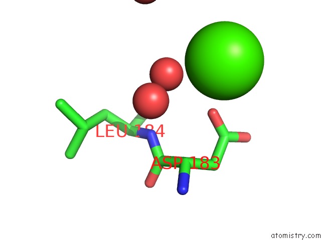
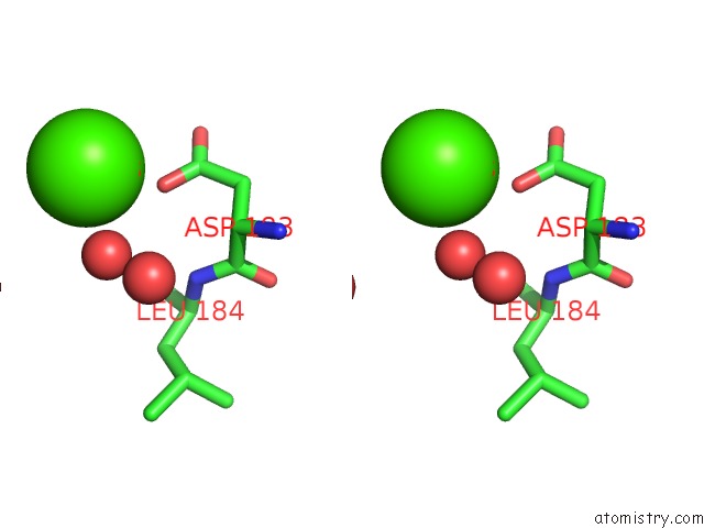
Calcium binding site 1 out of 3 in 2hrg
Go back to
Calcium binding site 1 out
of 3 in the Crystal Structure of Blue Laccase From Trametes Trogii Complexed with P-Methylbenzoate
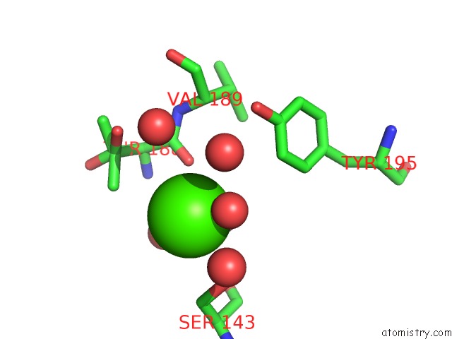
Mono view
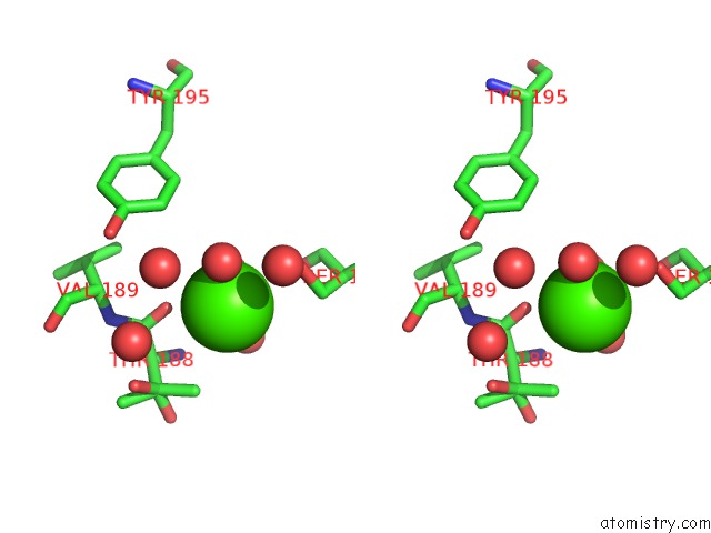
Stereo pair view
Mono view
Stereo pair view
A full contact list of Calcium with other atoms in the Ca binding
site number 1 of Crystal Structure of Blue Laccase From Trametes Trogii Complexed with P-Methylbenzoate within 5.0Å range:
|
Calcium binding site 2 out of 3 in 2hrg
Go back to
Calcium binding site 2 out
of 3 in the Crystal Structure of Blue Laccase From Trametes Trogii Complexed with P-Methylbenzoate
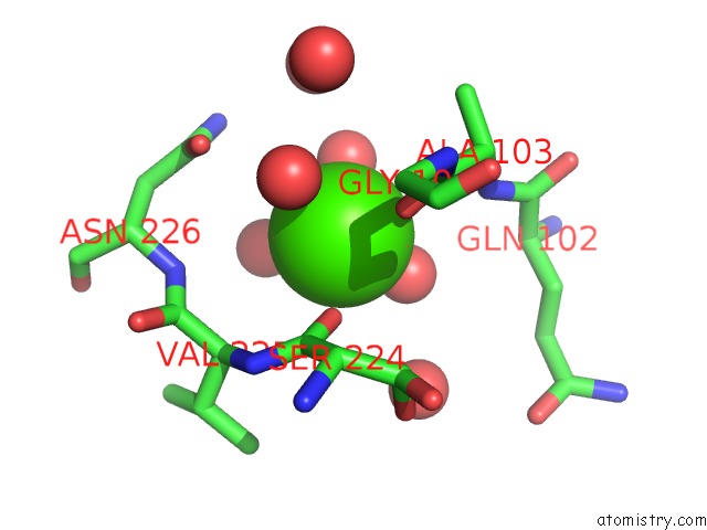
Mono view
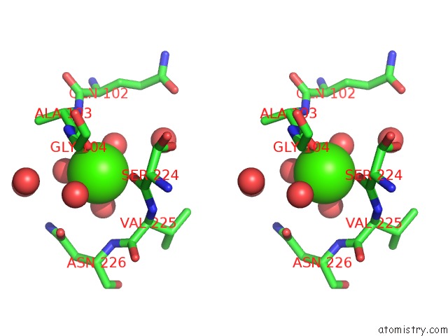
Stereo pair view
Mono view
Stereo pair view
A full contact list of Calcium with other atoms in the Ca binding
site number 2 of Crystal Structure of Blue Laccase From Trametes Trogii Complexed with P-Methylbenzoate within 5.0Å range:
|
Calcium binding site 3 out of 3 in 2hrg
Go back to
Calcium binding site 3 out
of 3 in the Crystal Structure of Blue Laccase From Trametes Trogii Complexed with P-Methylbenzoate
Mono view
Stereo pair view
Mono view
Stereo pair view
A full contact list of Calcium with other atoms in the Ca binding
site number 3 of Crystal Structure of Blue Laccase From Trametes Trogii Complexed with P-Methylbenzoate within 5.0Å range:
|
Reference:
I.Matera,
A.Gullotto,
S.Tilli,
M.Ferraroni,
A.Scozzafava,
F.Briganti.
Crystal Structure of the Blue Multicopper Oxidase From the White-Rot Fungus Trametes Trogii Complexed with P-Toluate Inorg.Chim.Acta. V. 361 4129 2008.
ISSN: ISSN 0020-1693
DOI: 10.1016/J.ICA.2008.03.091
Page generated: Fri Jul 12 12:48:39 2024
ISSN: ISSN 0020-1693
DOI: 10.1016/J.ICA.2008.03.091
Last articles
Zn in 9J0NZn in 9J0O
Zn in 9J0P
Zn in 9FJX
Zn in 9EKB
Zn in 9C0F
Zn in 9CAH
Zn in 9CH0
Zn in 9CH3
Zn in 9CH1