Calcium »
PDB 2i5w-2imw »
2i5w »
Calcium in PDB 2i5w: Structure of HOGG1 Crosslinked to Dna Sampling A Normal G Adjacent to An Oxog
Enzymatic activity of Structure of HOGG1 Crosslinked to Dna Sampling A Normal G Adjacent to An Oxog
All present enzymatic activity of Structure of HOGG1 Crosslinked to Dna Sampling A Normal G Adjacent to An Oxog:
4.2.99.18;
4.2.99.18;
Protein crystallography data
The structure of Structure of HOGG1 Crosslinked to Dna Sampling A Normal G Adjacent to An Oxog, PDB code: 2i5w
was solved by
A.Banerjee,
G.L.Verdine,
with X-Ray Crystallography technique. A brief refinement statistics is given in the table below:
| Resolution Low / High (Å) | 45.76 / 2.60 |
| Space group | P 65 2 2 |
| Cell size a, b, c (Å), α, β, γ (°) | 91.524, 91.524, 211.848, 90.00, 90.00, 120.00 |
| R / Rfree (%) | 22.6 / 26.6 |
Calcium Binding Sites:
The binding sites of Calcium atom in the Structure of HOGG1 Crosslinked to Dna Sampling A Normal G Adjacent to An Oxog
(pdb code 2i5w). This binding sites where shown within
5.0 Angstroms radius around Calcium atom.
In total 2 binding sites of Calcium where determined in the Structure of HOGG1 Crosslinked to Dna Sampling A Normal G Adjacent to An Oxog, PDB code: 2i5w:
Jump to Calcium binding site number: 1; 2;
In total 2 binding sites of Calcium where determined in the Structure of HOGG1 Crosslinked to Dna Sampling A Normal G Adjacent to An Oxog, PDB code: 2i5w:
Jump to Calcium binding site number: 1; 2;
Calcium binding site 1 out of 2 in 2i5w
Go back to
Calcium binding site 1 out
of 2 in the Structure of HOGG1 Crosslinked to Dna Sampling A Normal G Adjacent to An Oxog
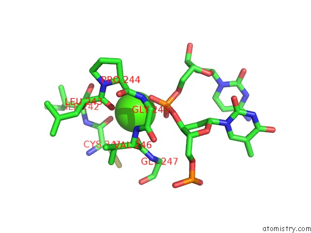
Mono view
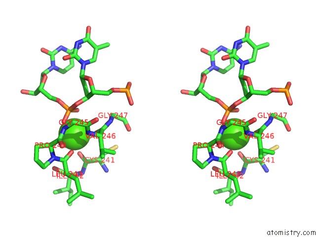
Stereo pair view
Mono view
Stereo pair view
A full contact list of Calcium with other atoms in the Ca binding
site number 1 of Structure of HOGG1 Crosslinked to Dna Sampling A Normal G Adjacent to An Oxog within 5.0Å range:
|
Calcium binding site 2 out of 2 in 2i5w
Go back to
Calcium binding site 2 out
of 2 in the Structure of HOGG1 Crosslinked to Dna Sampling A Normal G Adjacent to An Oxog
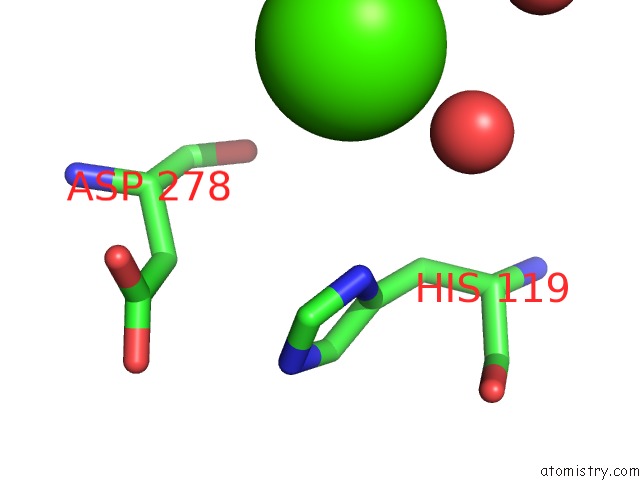
Mono view
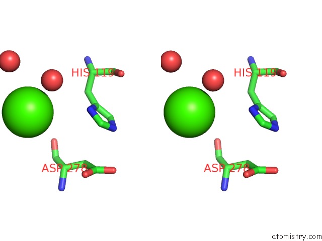
Stereo pair view
Mono view
Stereo pair view
A full contact list of Calcium with other atoms in the Ca binding
site number 2 of Structure of HOGG1 Crosslinked to Dna Sampling A Normal G Adjacent to An Oxog within 5.0Å range:
|
Reference:
A.Banerjee,
G.L.Verdine.
A Nucleobase Lesion Remodels the Interaction of Its Normal Neighbor in A Dna Glycosylase Complex. Proc.Natl.Acad.Sci.Usa V. 103 15020 2006.
ISSN: ISSN 0027-8424
PubMed: 17015827
DOI: 10.1073/PNAS.0603644103
Page generated: Tue Jul 8 06:10:33 2025
ISSN: ISSN 0027-8424
PubMed: 17015827
DOI: 10.1073/PNAS.0603644103
Last articles
Cl in 5L3XCl in 5L39
Cl in 5L38
Cl in 5L35
Cl in 5L2U
Cl in 5L2N
Cl in 5L1E
Cl in 5L2K
Cl in 5L0K
Cl in 5L2M