Calcium »
PDB 2i5w-2imw »
2iao »
Calcium in PDB 2iao: Crystal Structure of Squid Ganglion Dfpase E37Q Mutant
Enzymatic activity of Crystal Structure of Squid Ganglion Dfpase E37Q Mutant
All present enzymatic activity of Crystal Structure of Squid Ganglion Dfpase E37Q Mutant:
3.1.8.2;
3.1.8.2;
Protein crystallography data
The structure of Crystal Structure of Squid Ganglion Dfpase E37Q Mutant, PDB code: 2iao
was solved by
E.I.Scharff,
J.Koepke,
G.Fritzsch,
C.Luecke,
H.Rueterjans,
with X-Ray Crystallography technique. A brief refinement statistics is given in the table below:
| Resolution Low / High (Å) | 90.00 / 2.00 |
| Space group | P 21 21 21 |
| Cell size a, b, c (Å), α, β, γ (°) | 42.901, 81.696, 86.246, 90.00, 90.00, 90.00 |
| R / Rfree (%) | 19 / 24.9 |
Calcium Binding Sites:
The binding sites of Calcium atom in the Crystal Structure of Squid Ganglion Dfpase E37Q Mutant
(pdb code 2iao). This binding sites where shown within
5.0 Angstroms radius around Calcium atom.
In total 2 binding sites of Calcium where determined in the Crystal Structure of Squid Ganglion Dfpase E37Q Mutant, PDB code: 2iao:
Jump to Calcium binding site number: 1; 2;
In total 2 binding sites of Calcium where determined in the Crystal Structure of Squid Ganglion Dfpase E37Q Mutant, PDB code: 2iao:
Jump to Calcium binding site number: 1; 2;
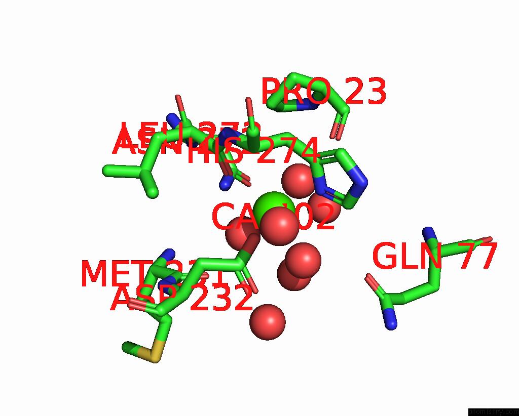
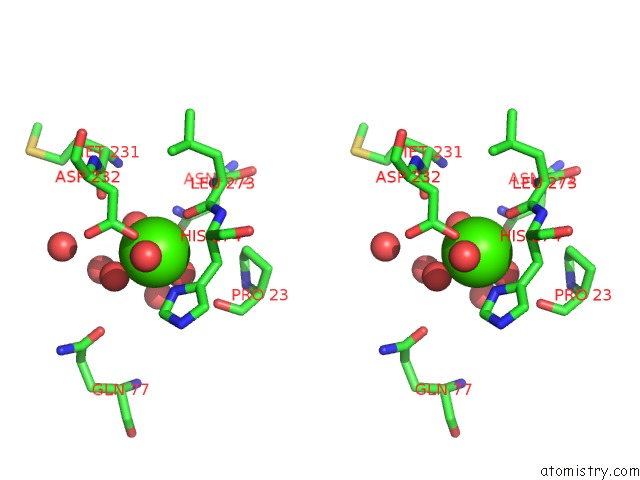
Calcium binding site 1 out of 2 in 2iao
Go back to
Calcium binding site 1 out
of 2 in the Crystal Structure of Squid Ganglion Dfpase E37Q Mutant

Mono view

Stereo pair view
Mono view
Stereo pair view
A full contact list of Calcium with other atoms in the Ca binding
site number 1 of Crystal Structure of Squid Ganglion Dfpase E37Q Mutant within 5.0Å range:
|
Calcium binding site 2 out of 2 in 2iao
Go back to
Calcium binding site 2 out
of 2 in the Crystal Structure of Squid Ganglion Dfpase E37Q Mutant
Mono view
Stereo pair view
Mono view
Stereo pair view
A full contact list of Calcium with other atoms in the Ca binding
site number 2 of Crystal Structure of Squid Ganglion Dfpase E37Q Mutant within 5.0Å range:
|
Reference:
E.I.Scharff,
J.Koepke,
G.Fritzsch,
C.Luecke,
H.Rueterjans.
Crystal Structure of Diisopropylfluorophosphatase From Loligo Vulgaris Structure V. 9 493 2001.
ISSN: ISSN 0969-2126
PubMed: 11435114
DOI: 10.1016/S0969-2126(01)00610-4
Page generated: Fri Jul 12 13:00:54 2024
ISSN: ISSN 0969-2126
PubMed: 11435114
DOI: 10.1016/S0969-2126(01)00610-4
Last articles
Zn in 9J0NZn in 9J0O
Zn in 9J0P
Zn in 9FJX
Zn in 9EKB
Zn in 9C0F
Zn in 9CAH
Zn in 9CH0
Zn in 9CH3
Zn in 9CH1