Calcium »
PDB 2i5w-2imw »
2iau »
Calcium in PDB 2iau: Crystal Structure of Squid Ganglion Dfpase W244Y Mutant
Enzymatic activity of Crystal Structure of Squid Ganglion Dfpase W244Y Mutant
All present enzymatic activity of Crystal Structure of Squid Ganglion Dfpase W244Y Mutant:
3.1.8.2;
3.1.8.2;
Protein crystallography data
The structure of Crystal Structure of Squid Ganglion Dfpase W244Y Mutant, PDB code: 2iau
was solved by
E.I.Scharff,
J.Koepke,
G.Fritzsch,
C.Luecke,
H.Rueterjans,
with X-Ray Crystallography technique. A brief refinement statistics is given in the table below:
| Resolution Low / High (Å) | 90.00 / 2.00 |
| Space group | P 21 21 21 |
| Cell size a, b, c (Å), α, β, γ (°) | 42.901, 81.696, 86.246, 90.00, 90.00, 90.00 |
| R / Rfree (%) | 20.9 / 26.4 |
Calcium Binding Sites:
The binding sites of Calcium atom in the Crystal Structure of Squid Ganglion Dfpase W244Y Mutant
(pdb code 2iau). This binding sites where shown within
5.0 Angstroms radius around Calcium atom.
In total 2 binding sites of Calcium where determined in the Crystal Structure of Squid Ganglion Dfpase W244Y Mutant, PDB code: 2iau:
Jump to Calcium binding site number: 1; 2;
In total 2 binding sites of Calcium where determined in the Crystal Structure of Squid Ganglion Dfpase W244Y Mutant, PDB code: 2iau:
Jump to Calcium binding site number: 1; 2;
Calcium binding site 1 out of 2 in 2iau
Go back to
Calcium binding site 1 out
of 2 in the Crystal Structure of Squid Ganglion Dfpase W244Y Mutant
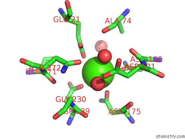
Mono view
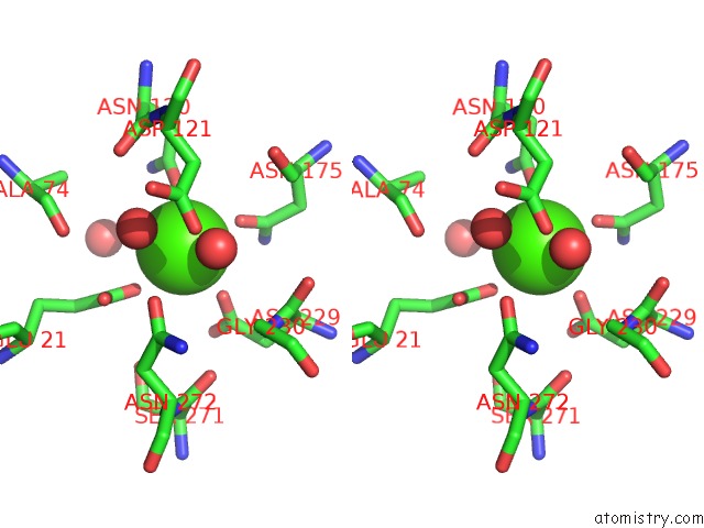
Stereo pair view
Mono view
Stereo pair view
A full contact list of Calcium with other atoms in the Ca binding
site number 1 of Crystal Structure of Squid Ganglion Dfpase W244Y Mutant within 5.0Å range:
|
Calcium binding site 2 out of 2 in 2iau
Go back to
Calcium binding site 2 out
of 2 in the Crystal Structure of Squid Ganglion Dfpase W244Y Mutant
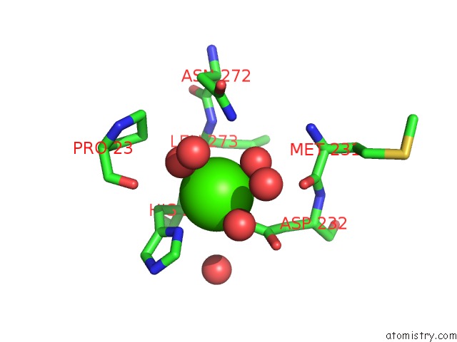
Mono view
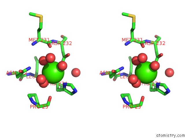
Stereo pair view
Mono view
Stereo pair view
A full contact list of Calcium with other atoms in the Ca binding
site number 2 of Crystal Structure of Squid Ganglion Dfpase W244Y Mutant within 5.0Å range:
|
Reference:
E.I.Scharff,
J.Koepke,
G.Fritzsch,
C.Luecke,
H.Rueterjans.
Crystal Structure of Diisopropylfluorophosphatase From Loligo Vulgaris Structure V. 9 493 2001.
ISSN: ISSN 0969-2126
PubMed: 11435114
DOI: 10.1016/S0969-2126(01)00610-4
Page generated: Tue Jul 8 06:12:16 2025
ISSN: ISSN 0969-2126
PubMed: 11435114
DOI: 10.1016/S0969-2126(01)00610-4
Last articles
F in 7MMJF in 7MMG
F in 7MMF
F in 7MMH
F in 7MMA
F in 7MM8
F in 7MM9
F in 7MHS
F in 7MII
F in 7MM7