Calcium »
PDB 2i5w-2imw »
2id4 »
Calcium in PDB 2id4: The 1.9 A Structure of KEX2 in Complex with An Ac-R-E-R-K-Chloromethyl Ketone Inhibitor.
Enzymatic activity of The 1.9 A Structure of KEX2 in Complex with An Ac-R-E-R-K-Chloromethyl Ketone Inhibitor.
All present enzymatic activity of The 1.9 A Structure of KEX2 in Complex with An Ac-R-E-R-K-Chloromethyl Ketone Inhibitor.:
3.4.21.61;
3.4.21.61;
Protein crystallography data
The structure of The 1.9 A Structure of KEX2 in Complex with An Ac-R-E-R-K-Chloromethyl Ketone Inhibitor., PDB code: 2id4
was solved by
J.L.Wheatley,
T.Holyoak,
with X-Ray Crystallography technique. A brief refinement statistics is given in the table below:
| Resolution Low / High (Å) | 40.79 / 1.90 |
| Space group | P 65 2 2 |
| Cell size a, b, c (Å), α, β, γ (°) | 112.851, 112.851, 370.165, 90.00, 90.00, 120.00 |
| R / Rfree (%) | 17.7 / 20.6 |
Other elements in 2id4:
The structure of The 1.9 A Structure of KEX2 in Complex with An Ac-R-E-R-K-Chloromethyl Ketone Inhibitor. also contains other interesting chemical elements:
| Sodium | (Na) | 2 atoms |
Calcium Binding Sites:
The binding sites of Calcium atom in the The 1.9 A Structure of KEX2 in Complex with An Ac-R-E-R-K-Chloromethyl Ketone Inhibitor.
(pdb code 2id4). This binding sites where shown within
5.0 Angstroms radius around Calcium atom.
In total 4 binding sites of Calcium where determined in the The 1.9 A Structure of KEX2 in Complex with An Ac-R-E-R-K-Chloromethyl Ketone Inhibitor., PDB code: 2id4:
Jump to Calcium binding site number: 1; 2; 3; 4;
In total 4 binding sites of Calcium where determined in the The 1.9 A Structure of KEX2 in Complex with An Ac-R-E-R-K-Chloromethyl Ketone Inhibitor., PDB code: 2id4:
Jump to Calcium binding site number: 1; 2; 3; 4;
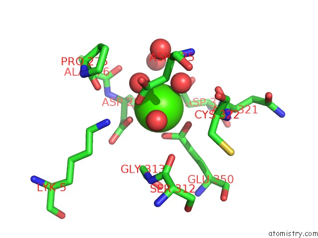
Calcium binding site 1 out of 4 in 2id4
Go back to
Calcium binding site 1 out
of 4 in the The 1.9 A Structure of KEX2 in Complex with An Ac-R-E-R-K-Chloromethyl Ketone Inhibitor.
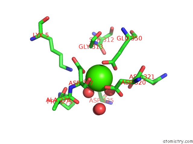
Mono view
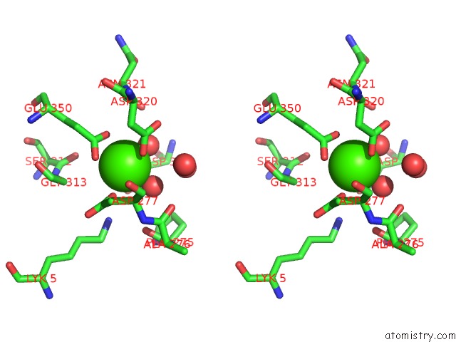
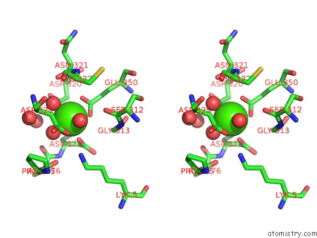
Stereo pair view
Mono view
Stereo pair view
A full contact list of Calcium with other atoms in the Ca binding
site number 1 of The 1.9 A Structure of KEX2 in Complex with An Ac-R-E-R-K-Chloromethyl Ketone Inhibitor. within 5.0Å range:
|
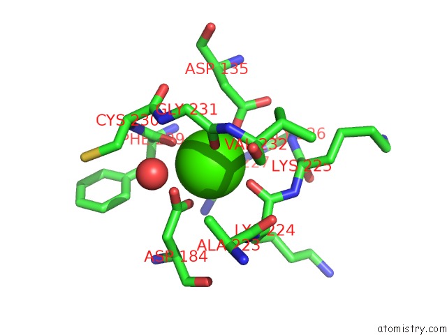
Calcium binding site 2 out of 4 in 2id4
Go back to
Calcium binding site 2 out
of 4 in the The 1.9 A Structure of KEX2 in Complex with An Ac-R-E-R-K-Chloromethyl Ketone Inhibitor.
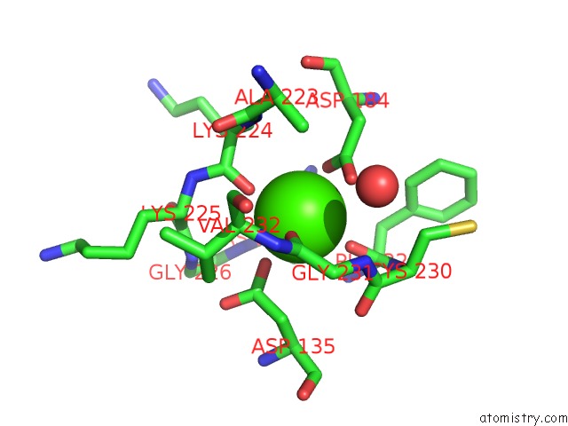
Mono view
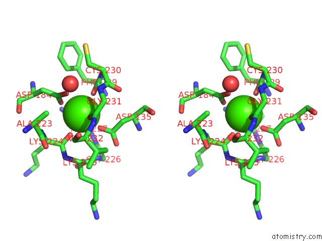
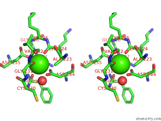
Stereo pair view
Mono view
Stereo pair view
A full contact list of Calcium with other atoms in the Ca binding
site number 2 of The 1.9 A Structure of KEX2 in Complex with An Ac-R-E-R-K-Chloromethyl Ketone Inhibitor. within 5.0Å range:
|
Calcium binding site 3 out of 4 in 2id4
Go back to
Calcium binding site 3 out
of 4 in the The 1.9 A Structure of KEX2 in Complex with An Ac-R-E-R-K-Chloromethyl Ketone Inhibitor.
Mono view
Stereo pair view
Mono view
Stereo pair view
A full contact list of Calcium with other atoms in the Ca binding
site number 3 of The 1.9 A Structure of KEX2 in Complex with An Ac-R-E-R-K-Chloromethyl Ketone Inhibitor. within 5.0Å range:
|
Calcium binding site 4 out of 4 in 2id4
Go back to
Calcium binding site 4 out
of 4 in the The 1.9 A Structure of KEX2 in Complex with An Ac-R-E-R-K-Chloromethyl Ketone Inhibitor.
Mono view
Stereo pair view
Mono view
Stereo pair view
A full contact list of Calcium with other atoms in the Ca binding
site number 4 of The 1.9 A Structure of KEX2 in Complex with An Ac-R-E-R-K-Chloromethyl Ketone Inhibitor. within 5.0Å range:
|
Reference:
J.L.Wheatley,
T.Holyoak.
Differential P1 Arginine and Lysine Recognition in the Prototypical Proprotein Convertase KEX2. Proc.Natl.Acad.Sci.Usa V. 104 6626 2007.
ISSN: ISSN 0027-8424
PubMed: 17426142
DOI: 10.1073/PNAS.0701983104
Page generated: Fri Jul 12 13:03:40 2024
ISSN: ISSN 0027-8424
PubMed: 17426142
DOI: 10.1073/PNAS.0701983104
Last articles
Zn in 9MJ5Zn in 9HNW
Zn in 9G0L
Zn in 9FNE
Zn in 9DZN
Zn in 9E0I
Zn in 9D32
Zn in 9DAK
Zn in 8ZXC
Zn in 8ZUF