Calcium »
PDB 2i5w-2imw »
2idj »
Calcium in PDB 2idj: Crystal Structure of Rat Glycine N-Methyltransferase Apoprotein, Monoclinic Form
Enzymatic activity of Crystal Structure of Rat Glycine N-Methyltransferase Apoprotein, Monoclinic Form
All present enzymatic activity of Crystal Structure of Rat Glycine N-Methyltransferase Apoprotein, Monoclinic Form:
2.1.1.20;
2.1.1.20;
Protein crystallography data
The structure of Crystal Structure of Rat Glycine N-Methyltransferase Apoprotein, Monoclinic Form, PDB code: 2idj
was solved by
Z.Luka,
S.Pakhomova,
L.V.Loukachevitch,
M.Egli,
M.E.Newcomer,
C.Wagner,
with X-Ray Crystallography technique. A brief refinement statistics is given in the table below:
| Resolution Low / High (Å) | 39.11 / 2.35 |
| Space group | P 1 21 1 |
| Cell size a, b, c (Å), α, β, γ (°) | 57.869, 85.223, 131.861, 90.00, 91.40, 90.00 |
| R / Rfree (%) | 22.2 / 27.2 |
Calcium Binding Sites:
The binding sites of Calcium atom in the Crystal Structure of Rat Glycine N-Methyltransferase Apoprotein, Monoclinic Form
(pdb code 2idj). This binding sites where shown within
5.0 Angstroms radius around Calcium atom.
In total 2 binding sites of Calcium where determined in the Crystal Structure of Rat Glycine N-Methyltransferase Apoprotein, Monoclinic Form, PDB code: 2idj:
Jump to Calcium binding site number: 1; 2;
In total 2 binding sites of Calcium where determined in the Crystal Structure of Rat Glycine N-Methyltransferase Apoprotein, Monoclinic Form, PDB code: 2idj:
Jump to Calcium binding site number: 1; 2;
Calcium binding site 1 out of 2 in 2idj
Go back to
Calcium binding site 1 out
of 2 in the Crystal Structure of Rat Glycine N-Methyltransferase Apoprotein, Monoclinic Form
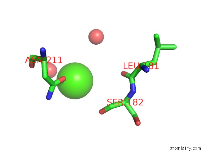
Mono view
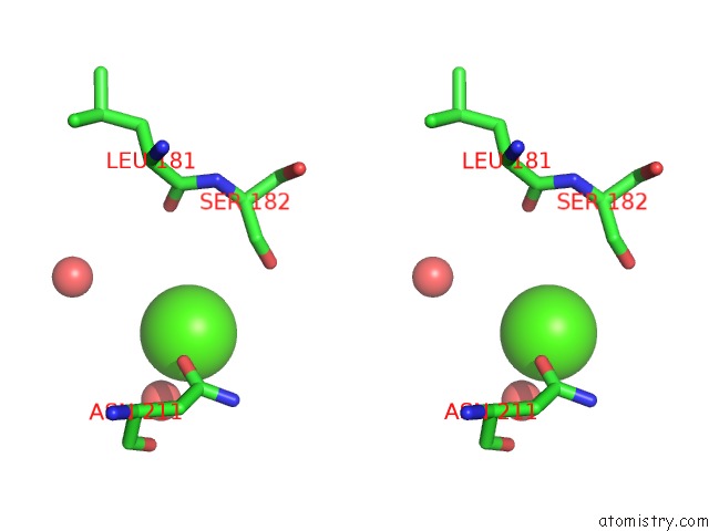
Stereo pair view
Mono view
Stereo pair view
A full contact list of Calcium with other atoms in the Ca binding
site number 1 of Crystal Structure of Rat Glycine N-Methyltransferase Apoprotein, Monoclinic Form within 5.0Å range:
|
Calcium binding site 2 out of 2 in 2idj
Go back to
Calcium binding site 2 out
of 2 in the Crystal Structure of Rat Glycine N-Methyltransferase Apoprotein, Monoclinic Form
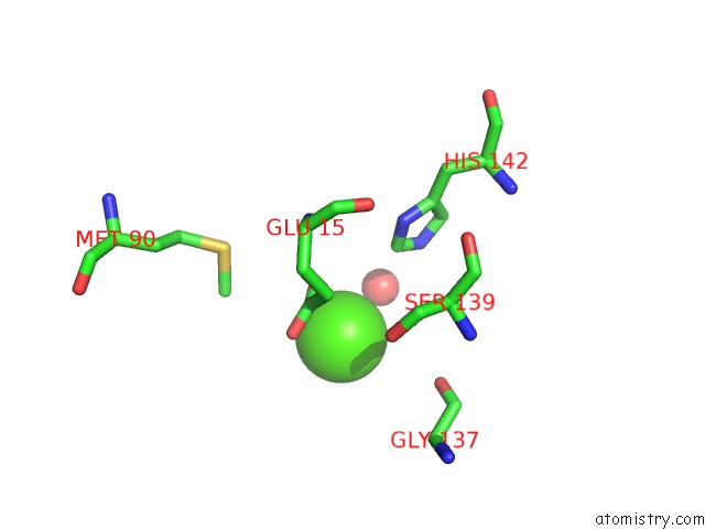
Mono view
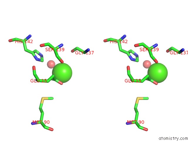
Stereo pair view
Mono view
Stereo pair view
A full contact list of Calcium with other atoms in the Ca binding
site number 2 of Crystal Structure of Rat Glycine N-Methyltransferase Apoprotein, Monoclinic Form within 5.0Å range:
|
Reference:
Z.Luka,
S.Pakhomova,
L.V.Loukachevitch,
M.Egli,
M.E.Newcomer,
C.Wagner.
5-Methyltetrahydrofolate Is Bound in Intersubunit Areas of Rat Liver Folate-Binding Protein Glycine N-Methyltransferase. J.Biol.Chem. V. 282 4069 2007.
ISSN: ISSN 0021-9258
PubMed: 17158459
DOI: 10.1074/JBC.M610384200
Page generated: Fri Jul 12 13:03:56 2024
ISSN: ISSN 0021-9258
PubMed: 17158459
DOI: 10.1074/JBC.M610384200
Last articles
Zn in 9MJ5Zn in 9HNW
Zn in 9G0L
Zn in 9FNE
Zn in 9DZN
Zn in 9E0I
Zn in 9D32
Zn in 9DAK
Zn in 8ZXC
Zn in 8ZUF