Calcium »
PDB 2io4-2j3o »
2it5 »
Calcium in PDB 2it5: Crystal Structure of Dcsign-Crd with MAN6
Protein crystallography data
The structure of Crystal Structure of Dcsign-Crd with MAN6, PDB code: 2it5
was solved by
W.I.Weis,
H.Feinberg,
R.Castelli,
K.Drickamer,
P.H.Seeberger,
with X-Ray Crystallography technique. A brief refinement statistics is given in the table below:
| Resolution Low / High (Å) | 27.98 / 2.40 |
| Space group | P 43 |
| Cell size a, b, c (Å), α, β, γ (°) | 55.960, 55.960, 53.260, 90.00, 90.00, 90.00 |
| R / Rfree (%) | 19.8 / 25.2 |
Calcium Binding Sites:
The binding sites of Calcium atom in the Crystal Structure of Dcsign-Crd with MAN6
(pdb code 2it5). This binding sites where shown within
5.0 Angstroms radius around Calcium atom.
In total 3 binding sites of Calcium where determined in the Crystal Structure of Dcsign-Crd with MAN6, PDB code: 2it5:
Jump to Calcium binding site number: 1; 2; 3;
In total 3 binding sites of Calcium where determined in the Crystal Structure of Dcsign-Crd with MAN6, PDB code: 2it5:
Jump to Calcium binding site number: 1; 2; 3;
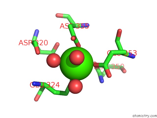
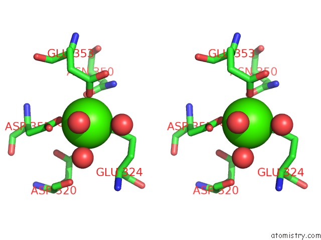
Calcium binding site 1 out of 3 in 2it5
Go back to
Calcium binding site 1 out
of 3 in the Crystal Structure of Dcsign-Crd with MAN6
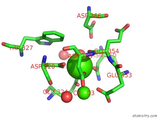
Mono view
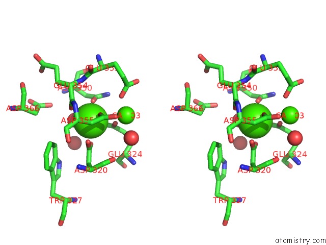
Stereo pair view
Mono view
Stereo pair view
A full contact list of Calcium with other atoms in the Ca binding
site number 1 of Crystal Structure of Dcsign-Crd with MAN6 within 5.0Å range:
|
Calcium binding site 2 out of 3 in 2it5
Go back to
Calcium binding site 2 out
of 3 in the Crystal Structure of Dcsign-Crd with MAN6
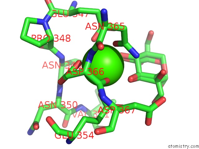
Mono view
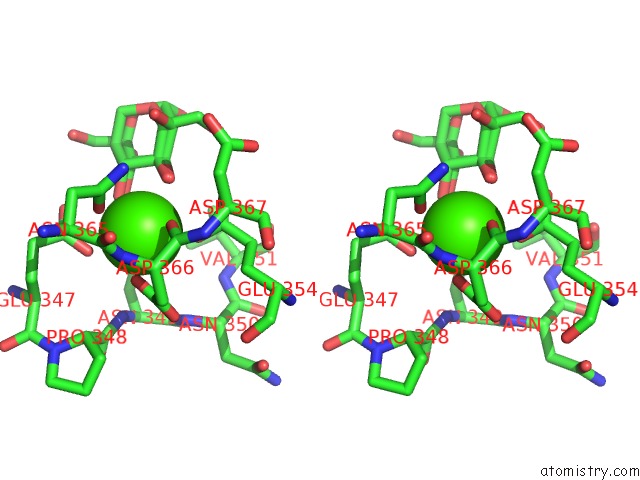
Stereo pair view
Mono view
Stereo pair view
A full contact list of Calcium with other atoms in the Ca binding
site number 2 of Crystal Structure of Dcsign-Crd with MAN6 within 5.0Å range:
|
Calcium binding site 3 out of 3 in 2it5
Go back to
Calcium binding site 3 out
of 3 in the Crystal Structure of Dcsign-Crd with MAN6
Mono view
Stereo pair view
Mono view
Stereo pair view
A full contact list of Calcium with other atoms in the Ca binding
site number 3 of Crystal Structure of Dcsign-Crd with MAN6 within 5.0Å range:
|
Reference:
H.Feinberg,
R.Castelli,
K.Drickamer,
P.H.Seeberger,
W.I.Weis.
Multiple Modes of Binding Enhance the Affinity of Dc-Sign For High Mannose N-Linked Glycans Found on Viral Glycoproteins. J.Biol.Chem. V. 282 4202 2007.
ISSN: ISSN 0021-9258
PubMed: 17150970
DOI: 10.1074/JBC.M609689200
Page generated: Tue Jul 8 06:17:17 2025
ISSN: ISSN 0021-9258
PubMed: 17150970
DOI: 10.1074/JBC.M609689200
Last articles
Cl in 5GRNCl in 5GSU
Cl in 5GS8
Cl in 5GQQ
Cl in 5G6U
Cl in 5GQN
Cl in 5GQM
Cl in 5GQL
Cl in 5GQK
Cl in 5GQJ