Calcium »
PDB 2io4-2j3o »
2j1e »
Calcium in PDB 2j1e: High Resolution Crystal Structure of CBM32 From A N-Acetyl- Beta-Hexosaminidase in Complex with Lacnac
Protein crystallography data
The structure of High Resolution Crystal Structure of CBM32 From A N-Acetyl- Beta-Hexosaminidase in Complex with Lacnac, PDB code: 2j1e
was solved by
E.Ficko-Blean,
A.B.Boraston,
with X-Ray Crystallography technique. A brief refinement statistics is given in the table below:
| Resolution Low / High (Å) | 20.00 / 2.4 |
| Space group | P 43 21 2 |
| Cell size a, b, c (Å), α, β, γ (°) | 77.377, 77.377, 73.421, 90.00, 90.00, 90.00 |
| R / Rfree (%) | 17.5 / 25.2 |
Calcium Binding Sites:
The binding sites of Calcium atom in the High Resolution Crystal Structure of CBM32 From A N-Acetyl- Beta-Hexosaminidase in Complex with Lacnac
(pdb code 2j1e). This binding sites where shown within
5.0 Angstroms radius around Calcium atom.
In total only one binding site of Calcium was determined in the High Resolution Crystal Structure of CBM32 From A N-Acetyl- Beta-Hexosaminidase in Complex with Lacnac, PDB code: 2j1e:
In total only one binding site of Calcium was determined in the High Resolution Crystal Structure of CBM32 From A N-Acetyl- Beta-Hexosaminidase in Complex with Lacnac, PDB code: 2j1e:
Calcium binding site 1 out of 1 in 2j1e
Go back to
Calcium binding site 1 out
of 1 in the High Resolution Crystal Structure of CBM32 From A N-Acetyl- Beta-Hexosaminidase in Complex with Lacnac
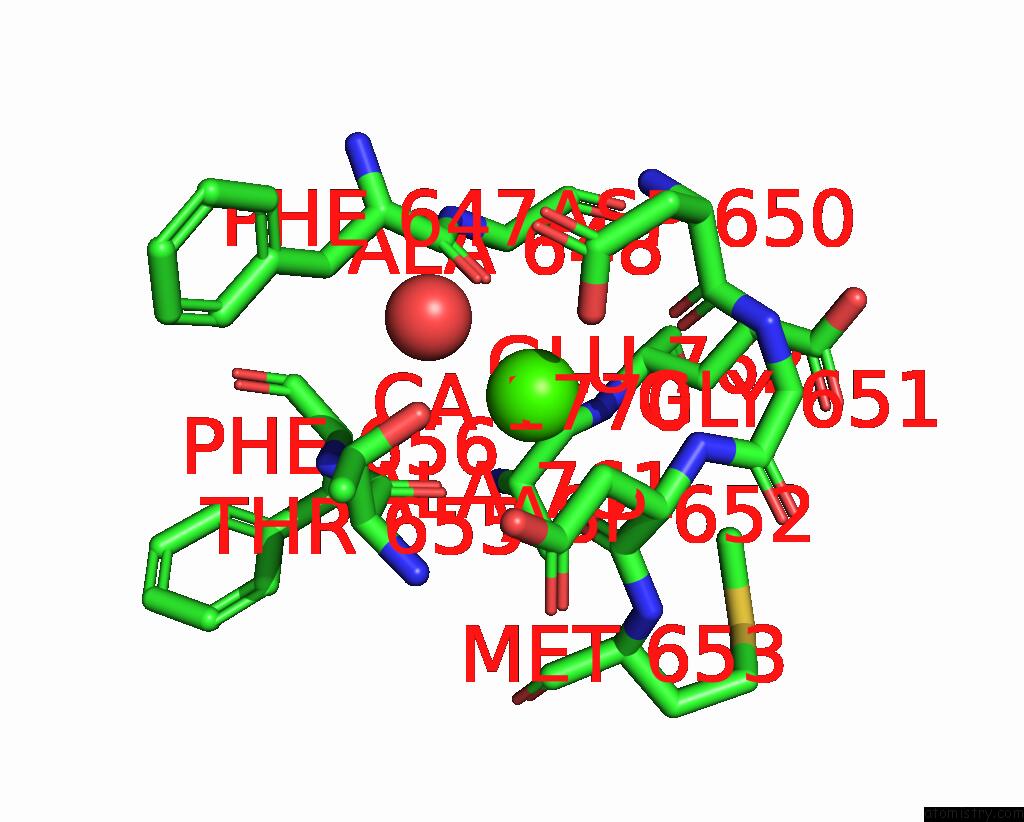
Mono view
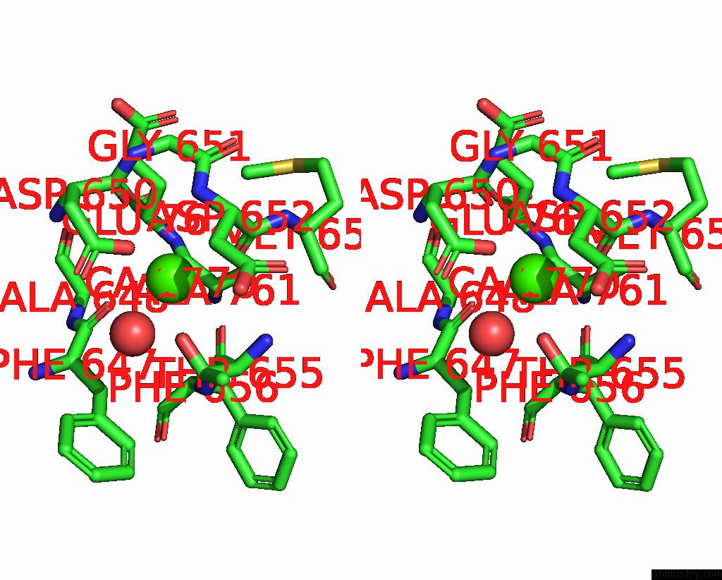
Stereo pair view
Mono view
Stereo pair view
A full contact list of Calcium with other atoms in the Ca binding
site number 1 of High Resolution Crystal Structure of CBM32 From A N-Acetyl- Beta-Hexosaminidase in Complex with Lacnac within 5.0Å range:
|
Reference:
E.Ficko-Blean,
A.B.Boraston.
The Interaction of A Carbohydrate-Binding Module From A Clostridium Perfringens N-Acetyl-Beta- Hexosaminidase with Its Carbohydrate Receptor J.Biol.Chem. V. 281 37748 2006.
ISSN: ISSN 0021-9258
PubMed: 16990278
DOI: 10.1074/JBC.M606126200
Page generated: Tue Jul 8 06:25:19 2025
ISSN: ISSN 0021-9258
PubMed: 16990278
DOI: 10.1074/JBC.M606126200
Last articles
Cl in 5GWBCl in 5GWA
Cl in 5GVP
Cl in 5GVN
Cl in 5GVM
Cl in 5GTY
Cl in 5GVL
Cl in 5GVK
Cl in 5GUS
Cl in 5GUW