Calcium »
PDB 2j3u-2jdh »
2j7t »
Calcium in PDB 2j7t: Crystal Structure of Human Serine Threonine Kinase-10 Bound to SU11274
Enzymatic activity of Crystal Structure of Human Serine Threonine Kinase-10 Bound to SU11274
All present enzymatic activity of Crystal Structure of Human Serine Threonine Kinase-10 Bound to SU11274:
2.7.11.1;
2.7.11.1;
Protein crystallography data
The structure of Crystal Structure of Human Serine Threonine Kinase-10 Bound to SU11274, PDB code: 2j7t
was solved by
A.C.W.Pike,
P.Rellos,
O.Fedorov,
S.Das,
J.Debreczeni,
F.Sobott,
S.Watt,
P.Savitsky,
J.Eswaran,
A.P.Turnbull,
E.Papagrigoriou,
E.Ugochukwa,
F.Gorrec,
C.C.Umeano,
F.Von Delft,
C.H.Arrowsmith,
A.Edwards,
J.Weigelt,
M.Sundstrom,
S.Knapp,
with X-Ray Crystallography technique. A brief refinement statistics is given in the table below:
| Resolution Low / High (Å) | 55.00 / 2.00 |
| Space group | I 2 2 2 |
| Cell size a, b, c (Å), α, β, γ (°) | 49.192, 112.960, 133.817, 90.00, 90.00, 90.00 |
| R / Rfree (%) | 20.4 / 23.5 |
Other elements in 2j7t:
The structure of Crystal Structure of Human Serine Threonine Kinase-10 Bound to SU11274 also contains other interesting chemical elements:
| Chlorine | (Cl) | 1 atom |
Calcium Binding Sites:
The binding sites of Calcium atom in the Crystal Structure of Human Serine Threonine Kinase-10 Bound to SU11274
(pdb code 2j7t). This binding sites where shown within
5.0 Angstroms radius around Calcium atom.
In total 7 binding sites of Calcium where determined in the Crystal Structure of Human Serine Threonine Kinase-10 Bound to SU11274, PDB code: 2j7t:
Jump to Calcium binding site number: 1; 2; 3; 4; 5; 6; 7;
In total 7 binding sites of Calcium where determined in the Crystal Structure of Human Serine Threonine Kinase-10 Bound to SU11274, PDB code: 2j7t:
Jump to Calcium binding site number: 1; 2; 3; 4; 5; 6; 7;
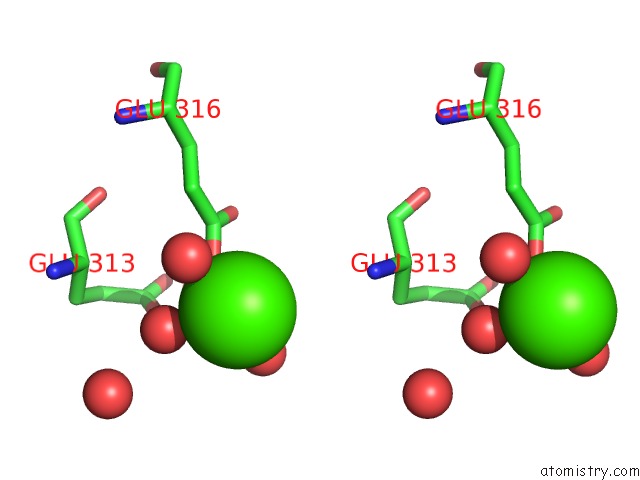
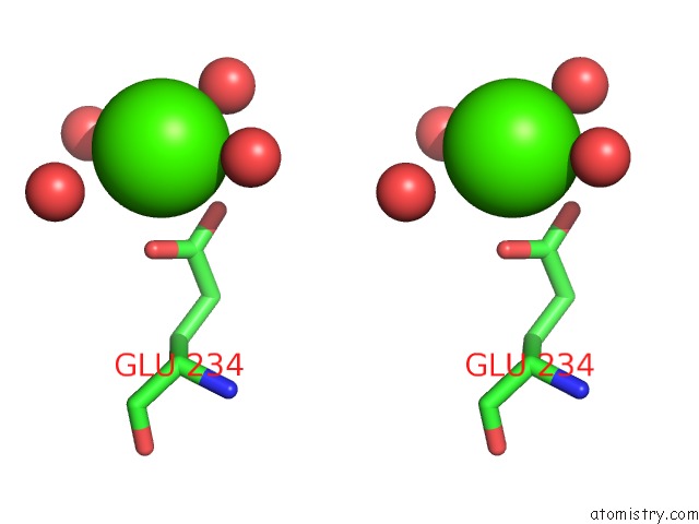
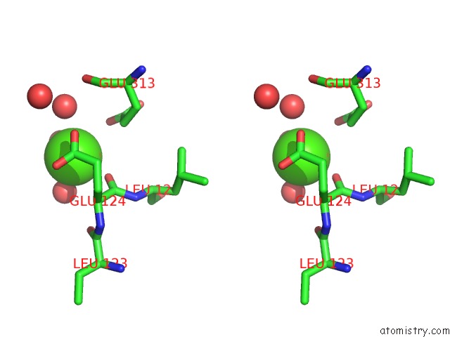
Calcium binding site 1 out of 7 in 2j7t
Go back to
Calcium binding site 1 out
of 7 in the Crystal Structure of Human Serine Threonine Kinase-10 Bound to SU11274
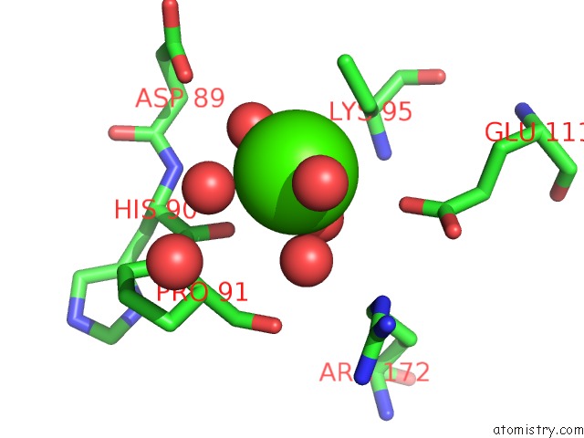
Mono view
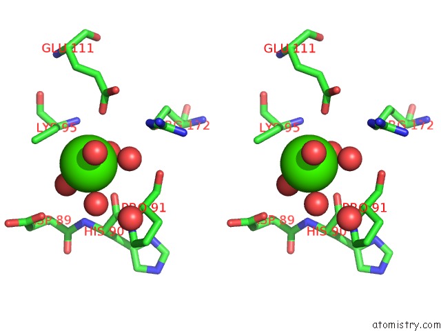
Stereo pair view
Mono view
Stereo pair view
A full contact list of Calcium with other atoms in the Ca binding
site number 1 of Crystal Structure of Human Serine Threonine Kinase-10 Bound to SU11274 within 5.0Å range:
|
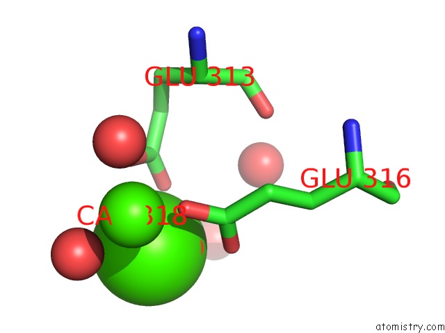
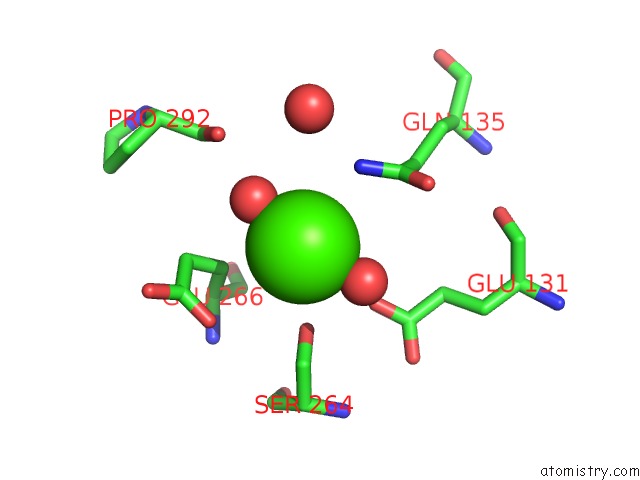
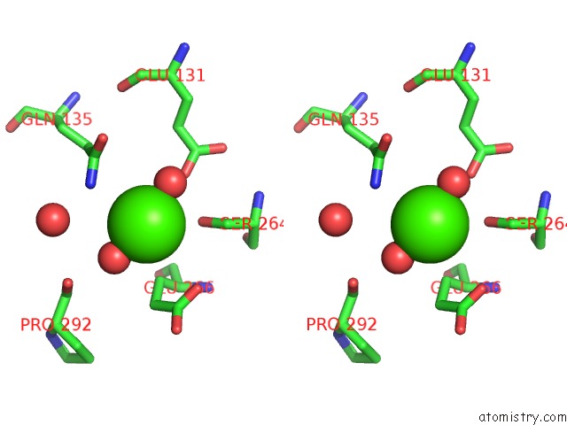
Calcium binding site 2 out of 7 in 2j7t
Go back to
Calcium binding site 2 out
of 7 in the Crystal Structure of Human Serine Threonine Kinase-10 Bound to SU11274
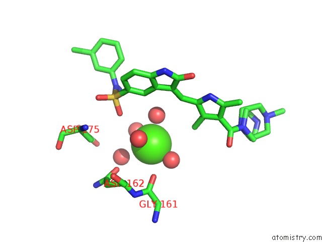
Mono view
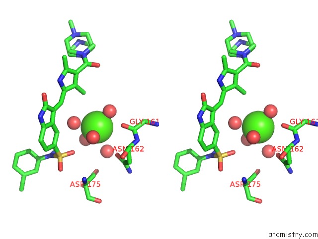
Stereo pair view
Mono view
Stereo pair view
A full contact list of Calcium with other atoms in the Ca binding
site number 2 of Crystal Structure of Human Serine Threonine Kinase-10 Bound to SU11274 within 5.0Å range:
|
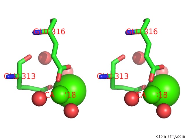
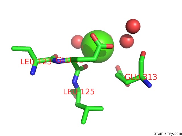
Calcium binding site 3 out of 7 in 2j7t
Go back to
Calcium binding site 3 out
of 7 in the Crystal Structure of Human Serine Threonine Kinase-10 Bound to SU11274

Mono view
Stereo pair view
Mono view
Stereo pair view
A full contact list of Calcium with other atoms in the Ca binding
site number 3 of Crystal Structure of Human Serine Threonine Kinase-10 Bound to SU11274 within 5.0Å range:
|
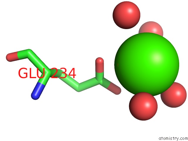
Calcium binding site 4 out of 7 in 2j7t
Go back to
Calcium binding site 4 out
of 7 in the Crystal Structure of Human Serine Threonine Kinase-10 Bound to SU11274
Mono view
Stereo pair view
Mono view
Stereo pair view
A full contact list of Calcium with other atoms in the Ca binding
site number 4 of Crystal Structure of Human Serine Threonine Kinase-10 Bound to SU11274 within 5.0Å range:
|
Calcium binding site 5 out of 7 in 2j7t
Go back to
Calcium binding site 5 out
of 7 in the Crystal Structure of Human Serine Threonine Kinase-10 Bound to SU11274
Mono view
Stereo pair view
Mono view
Stereo pair view
A full contact list of Calcium with other atoms in the Ca binding
site number 5 of Crystal Structure of Human Serine Threonine Kinase-10 Bound to SU11274 within 5.0Å range:
|
Calcium binding site 6 out of 7 in 2j7t
Go back to
Calcium binding site 6 out
of 7 in the Crystal Structure of Human Serine Threonine Kinase-10 Bound to SU11274
Mono view
Stereo pair view
Mono view
Stereo pair view
A full contact list of Calcium with other atoms in the Ca binding
site number 6 of Crystal Structure of Human Serine Threonine Kinase-10 Bound to SU11274 within 5.0Å range:
|
Calcium binding site 7 out of 7 in 2j7t
Go back to
Calcium binding site 7 out
of 7 in the Crystal Structure of Human Serine Threonine Kinase-10 Bound to SU11274
Mono view
Stereo pair view
Mono view
Stereo pair view
A full contact list of Calcium with other atoms in the Ca binding
site number 7 of Crystal Structure of Human Serine Threonine Kinase-10 Bound to SU11274 within 5.0Å range:
|
Reference:
A.C.W.Pike,
P.Rellos,
F.H.Niesen,
A.Turnbull,
A.W.Oliver,
S.A.Parker,
B.E.Turk,
L.H.Pearl,
S.Knapp.
Activation Segment Dimerization: A Mechanism For Kinase Autophosphorylation of Non-Consensus Sites. Embo J. V. 27 704 2008.
ISSN: ISSN 0261-4189
PubMed: 18239682
DOI: 10.1038/EMBOJ.2008.8
Page generated: Fri Jul 12 13:31:27 2024
ISSN: ISSN 0261-4189
PubMed: 18239682
DOI: 10.1038/EMBOJ.2008.8
Last articles
Zn in 9J0NZn in 9J0O
Zn in 9J0P
Zn in 9FJX
Zn in 9EKB
Zn in 9C0F
Zn in 9CAH
Zn in 9CH0
Zn in 9CH3
Zn in 9CH1