Calcium »
PDB 2j3u-2jdh »
2jda »
Calcium in PDB 2jda: Structure of A Pectin Binding Carbohydrate Binding Module Determined in An Monoclinic Crystal Form.
Protein crystallography data
The structure of Structure of A Pectin Binding Carbohydrate Binding Module Determined in An Monoclinic Crystal Form., PDB code: 2jda
was solved by
D.W.Abbott,
A.B.Boraston,
with X-Ray Crystallography technique. A brief refinement statistics is given in the table below:
| Resolution Low / High (Å) | 20.00 / 1.35 |
| Space group | C 1 2 1 |
| Cell size a, b, c (Å), α, β, γ (°) | 111.205, 34.729, 86.711, 90.00, 120.58, 90.00 |
| R / Rfree (%) | 14.3 / 18 |
Calcium Binding Sites:
The binding sites of Calcium atom in the Structure of A Pectin Binding Carbohydrate Binding Module Determined in An Monoclinic Crystal Form.
(pdb code 2jda). This binding sites where shown within
5.0 Angstroms radius around Calcium atom.
In total 2 binding sites of Calcium where determined in the Structure of A Pectin Binding Carbohydrate Binding Module Determined in An Monoclinic Crystal Form., PDB code: 2jda:
Jump to Calcium binding site number: 1; 2;
In total 2 binding sites of Calcium where determined in the Structure of A Pectin Binding Carbohydrate Binding Module Determined in An Monoclinic Crystal Form., PDB code: 2jda:
Jump to Calcium binding site number: 1; 2;
Calcium binding site 1 out of 2 in 2jda
Go back to
Calcium binding site 1 out
of 2 in the Structure of A Pectin Binding Carbohydrate Binding Module Determined in An Monoclinic Crystal Form.
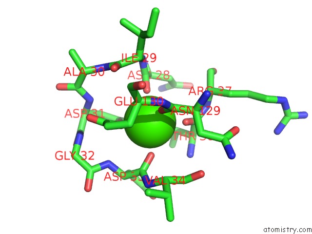
Mono view
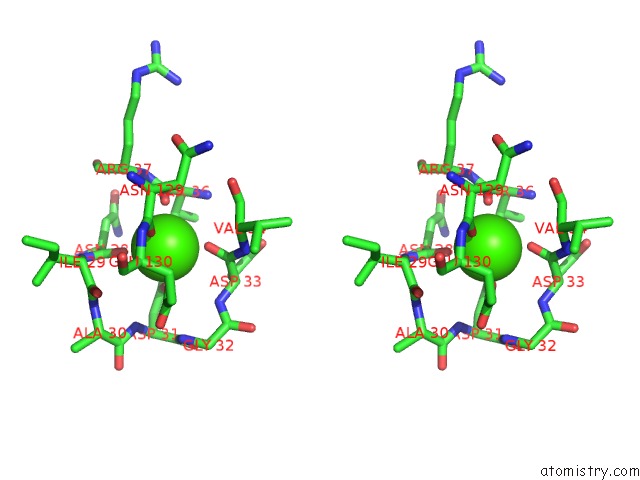
Stereo pair view
Mono view
Stereo pair view
A full contact list of Calcium with other atoms in the Ca binding
site number 1 of Structure of A Pectin Binding Carbohydrate Binding Module Determined in An Monoclinic Crystal Form. within 5.0Å range:
|
Calcium binding site 2 out of 2 in 2jda
Go back to
Calcium binding site 2 out
of 2 in the Structure of A Pectin Binding Carbohydrate Binding Module Determined in An Monoclinic Crystal Form.
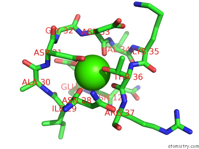
Mono view
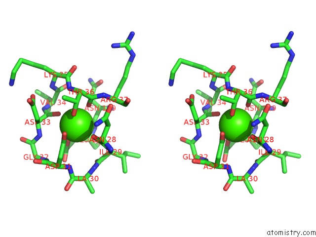
Stereo pair view
Mono view
Stereo pair view
A full contact list of Calcium with other atoms in the Ca binding
site number 2 of Structure of A Pectin Binding Carbohydrate Binding Module Determined in An Monoclinic Crystal Form. within 5.0Å range:
|
Reference:
D.W.Abbott,
S.Hrynuik,
A.B.Boraston.
Identification and Characterization of A Novel Periplasmic Polygalacturonic Acid Binding Protein From Yersinia Enterolitica J.Mol.Biol. V. 367 1023 2007.
ISSN: ISSN 0022-2836
PubMed: 17292916
DOI: 10.1016/J.JMB.2007.01.030
Page generated: Fri Jul 12 13:34:10 2024
ISSN: ISSN 0022-2836
PubMed: 17292916
DOI: 10.1016/J.JMB.2007.01.030
Last articles
Zn in 9J0NZn in 9J0O
Zn in 9J0P
Zn in 9FJX
Zn in 9EKB
Zn in 9C0F
Zn in 9CAH
Zn in 9CH0
Zn in 9CH3
Zn in 9CH1