Calcium »
PDB 2jdk-2jnx »
2ji2 »
Calcium in PDB 2ji2: X-Ray Structure of E114A Mutant of Superoxide Reductase From Desulfoarculus Baarsii in the Native, Reduced Form
Enzymatic activity of X-Ray Structure of E114A Mutant of Superoxide Reductase From Desulfoarculus Baarsii in the Native, Reduced Form
All present enzymatic activity of X-Ray Structure of E114A Mutant of Superoxide Reductase From Desulfoarculus Baarsii in the Native, Reduced Form:
1.15.1.2;
1.15.1.2;
Protein crystallography data
The structure of X-Ray Structure of E114A Mutant of Superoxide Reductase From Desulfoarculus Baarsii in the Native, Reduced Form, PDB code: 2ji2
was solved by
G.Katona,
P.Carpentier,
V.Niviere,
P.Amara,
V.Adam,
J.Ohana,
N.Tsanov,
D.Bourgeois,
with X-Ray Crystallography technique. A brief refinement statistics is given in the table below:
| Resolution Low / High (Å) | 47.40 / 1.70 |
| Space group | I 2 2 2 |
| Cell size a, b, c (Å), α, β, γ (°) | 70.470, 82.870, 201.950, 90.00, 90.00, 90.00 |
| R / Rfree (%) | 19.5 / 22.2 |
Other elements in 2ji2:
The structure of X-Ray Structure of E114A Mutant of Superoxide Reductase From Desulfoarculus Baarsii in the Native, Reduced Form also contains other interesting chemical elements:
| Iron | (Fe) | 8 atoms |
Calcium Binding Sites:
The binding sites of Calcium atom in the X-Ray Structure of E114A Mutant of Superoxide Reductase From Desulfoarculus Baarsii in the Native, Reduced Form
(pdb code 2ji2). This binding sites where shown within
5.0 Angstroms radius around Calcium atom.
In total 4 binding sites of Calcium where determined in the X-Ray Structure of E114A Mutant of Superoxide Reductase From Desulfoarculus Baarsii in the Native, Reduced Form, PDB code: 2ji2:
Jump to Calcium binding site number: 1; 2; 3; 4;
In total 4 binding sites of Calcium where determined in the X-Ray Structure of E114A Mutant of Superoxide Reductase From Desulfoarculus Baarsii in the Native, Reduced Form, PDB code: 2ji2:
Jump to Calcium binding site number: 1; 2; 3; 4;
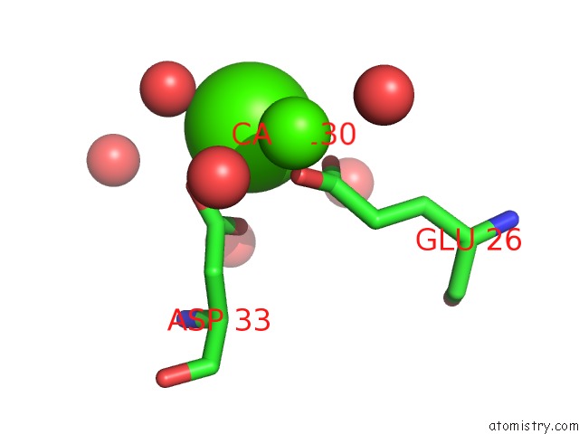
Calcium binding site 1 out of 4 in 2ji2
Go back to
Calcium binding site 1 out
of 4 in the X-Ray Structure of E114A Mutant of Superoxide Reductase From Desulfoarculus Baarsii in the Native, Reduced Form
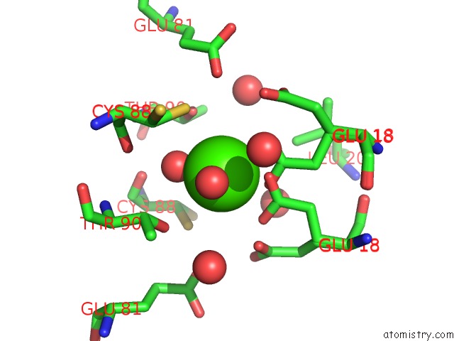
Mono view
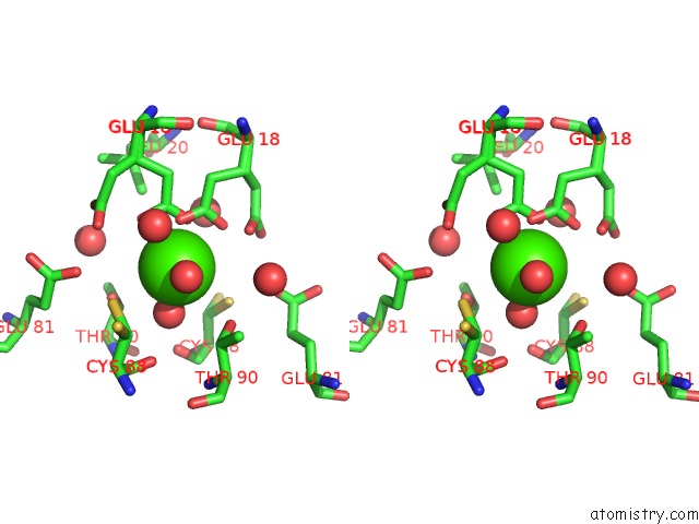
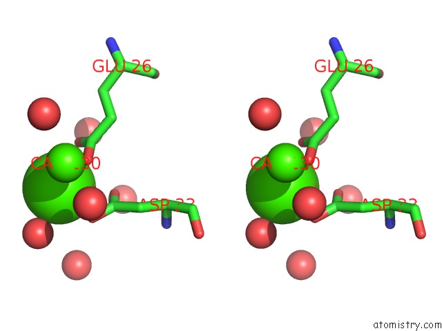
Stereo pair view
Mono view
Stereo pair view
A full contact list of Calcium with other atoms in the Ca binding
site number 1 of X-Ray Structure of E114A Mutant of Superoxide Reductase From Desulfoarculus Baarsii in the Native, Reduced Form within 5.0Å range:
|
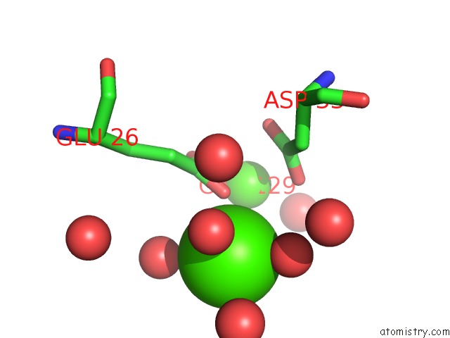
Calcium binding site 2 out of 4 in 2ji2
Go back to
Calcium binding site 2 out
of 4 in the X-Ray Structure of E114A Mutant of Superoxide Reductase From Desulfoarculus Baarsii in the Native, Reduced Form
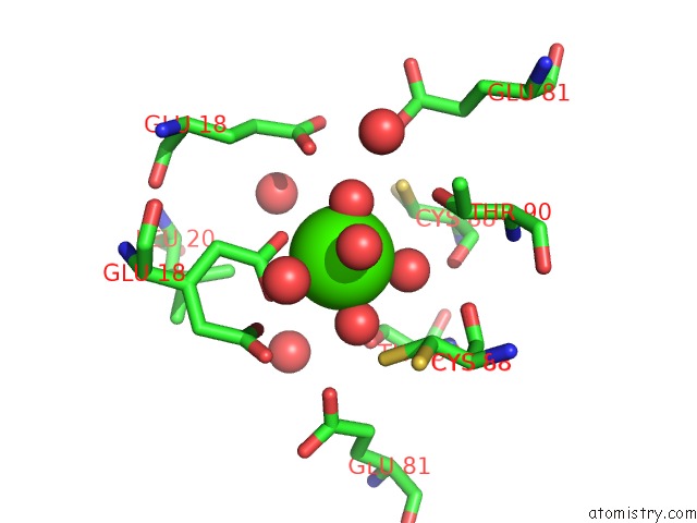
Mono view
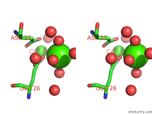
Stereo pair view
Mono view
Stereo pair view
A full contact list of Calcium with other atoms in the Ca binding
site number 2 of X-Ray Structure of E114A Mutant of Superoxide Reductase From Desulfoarculus Baarsii in the Native, Reduced Form within 5.0Å range:
|
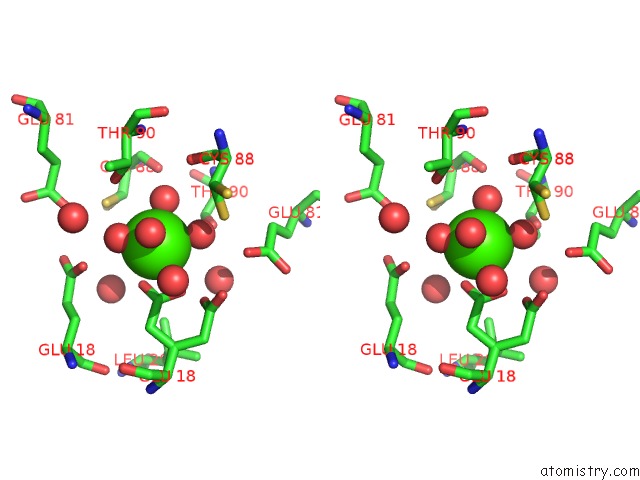
Calcium binding site 3 out of 4 in 2ji2
Go back to
Calcium binding site 3 out
of 4 in the X-Ray Structure of E114A Mutant of Superoxide Reductase From Desulfoarculus Baarsii in the Native, Reduced Form
Mono view
Stereo pair view
Mono view
Stereo pair view
A full contact list of Calcium with other atoms in the Ca binding
site number 3 of X-Ray Structure of E114A Mutant of Superoxide Reductase From Desulfoarculus Baarsii in the Native, Reduced Form within 5.0Å range:
|
Calcium binding site 4 out of 4 in 2ji2
Go back to
Calcium binding site 4 out
of 4 in the X-Ray Structure of E114A Mutant of Superoxide Reductase From Desulfoarculus Baarsii in the Native, Reduced Form
Mono view
Stereo pair view
Mono view
Stereo pair view
A full contact list of Calcium with other atoms in the Ca binding
site number 4 of X-Ray Structure of E114A Mutant of Superoxide Reductase From Desulfoarculus Baarsii in the Native, Reduced Form within 5.0Å range:
|
Reference:
G.Katona,
P.Carpentier,
V.Niviere,
P.Amara,
V.Adam,
J.Ohana,
N.Tsanov,
D.Bourgeois.
Raman-Assisted Crystallography Reveals End-on Peroxide Intermediates in A Nonheme Iron Enzyme. Science V. 316 449 2007.
ISSN: ESSN 1095-9203
PubMed: 17446401
DOI: 10.1126/SCIENCE.1138885
Page generated: Tue Jul 8 06:43:00 2025
ISSN: ESSN 1095-9203
PubMed: 17446401
DOI: 10.1126/SCIENCE.1138885
Last articles
Cl in 5J95Cl in 5J7G
Cl in 5J8Z
Cl in 5J8F
Cl in 5J8I
Cl in 5J8C
Cl in 5J7S
Cl in 5J7R
Cl in 5J7F
Cl in 5J72