Calcium »
PDB 2jdk-2jnx »
2jnx »
Calcium in PDB 2jnx: uc(Nmr) Derived Solution Structure of An Ef-Hand Calcium Binding Protein From Entamoeba Histolytica
Calcium Binding Sites:
The binding sites of Calcium atom in the uc(Nmr) Derived Solution Structure of An Ef-Hand Calcium Binding Protein From Entamoeba Histolytica
(pdb code 2jnx). This binding sites where shown within
5.0 Angstroms radius around Calcium atom.
In total 4 binding sites of Calcium where determined in the uc(Nmr) Derived Solution Structure of An Ef-Hand Calcium Binding Protein From Entamoeba Histolytica, PDB code: 2jnx:
Jump to Calcium binding site number: 1; 2; 3; 4;
In total 4 binding sites of Calcium where determined in the uc(Nmr) Derived Solution Structure of An Ef-Hand Calcium Binding Protein From Entamoeba Histolytica, PDB code: 2jnx:
Jump to Calcium binding site number: 1; 2; 3; 4;
Calcium binding site 1 out of 4 in 2jnx
Go back to
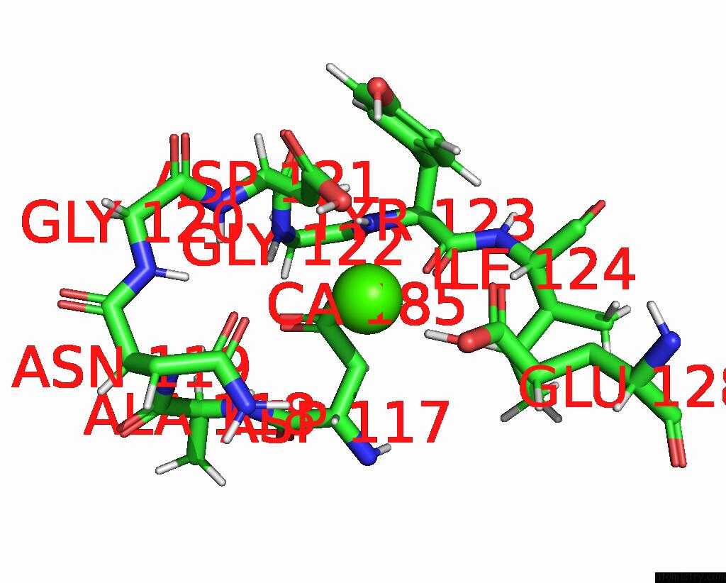
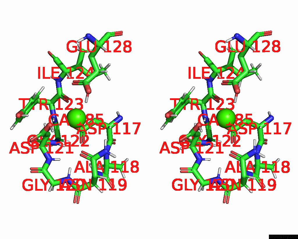
Calcium binding site 1 out
of 4 in the uc(Nmr) Derived Solution Structure of An Ef-Hand Calcium Binding Protein From Entamoeba Histolytica
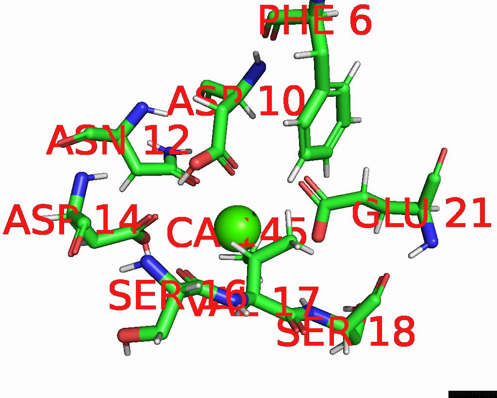
Mono view

Stereo pair view
Mono view
Stereo pair view
A full contact list of Calcium with other atoms in the Ca binding
site number 1 of uc(Nmr) Derived Solution Structure of An Ef-Hand Calcium Binding Protein From Entamoeba Histolytica within 5.0Å range:
|
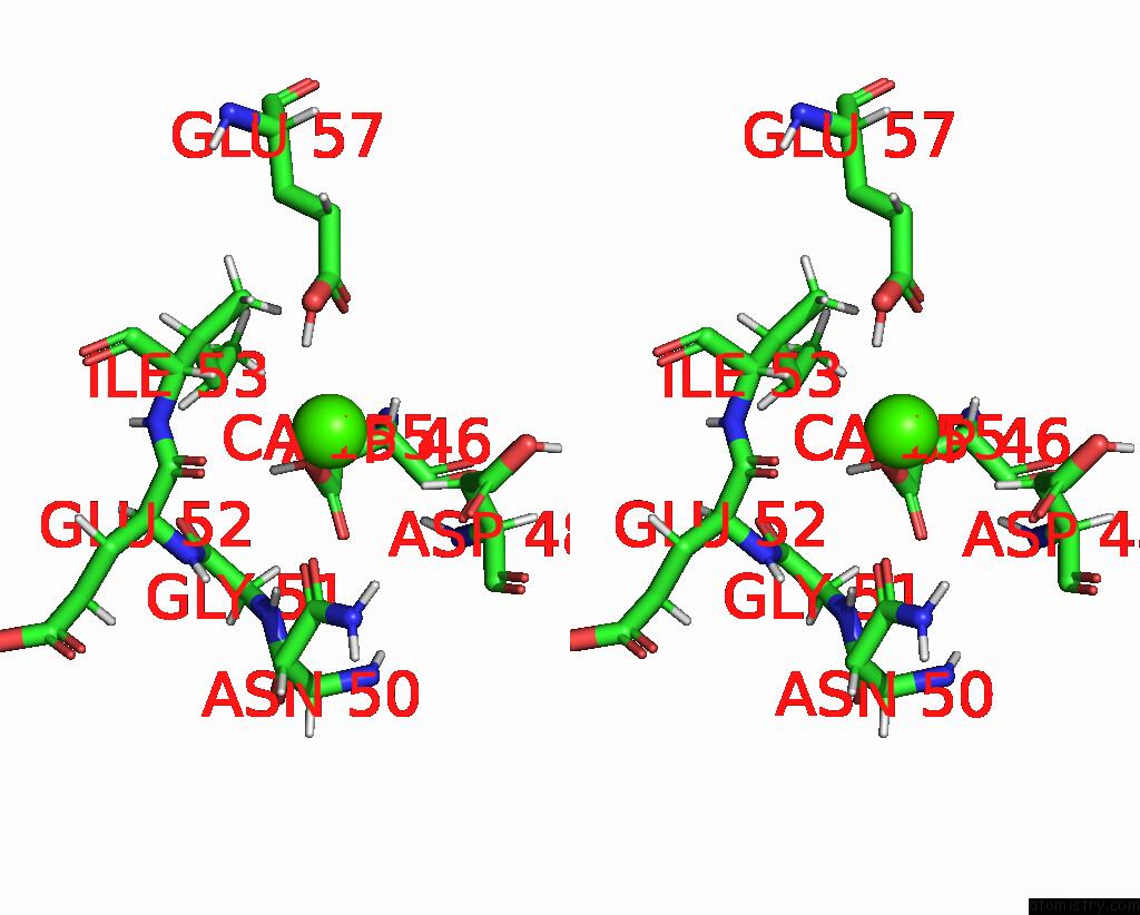
Calcium binding site 2 out of 4 in 2jnx
Go back to
Calcium binding site 2 out
of 4 in the uc(Nmr) Derived Solution Structure of An Ef-Hand Calcium Binding Protein From Entamoeba Histolytica
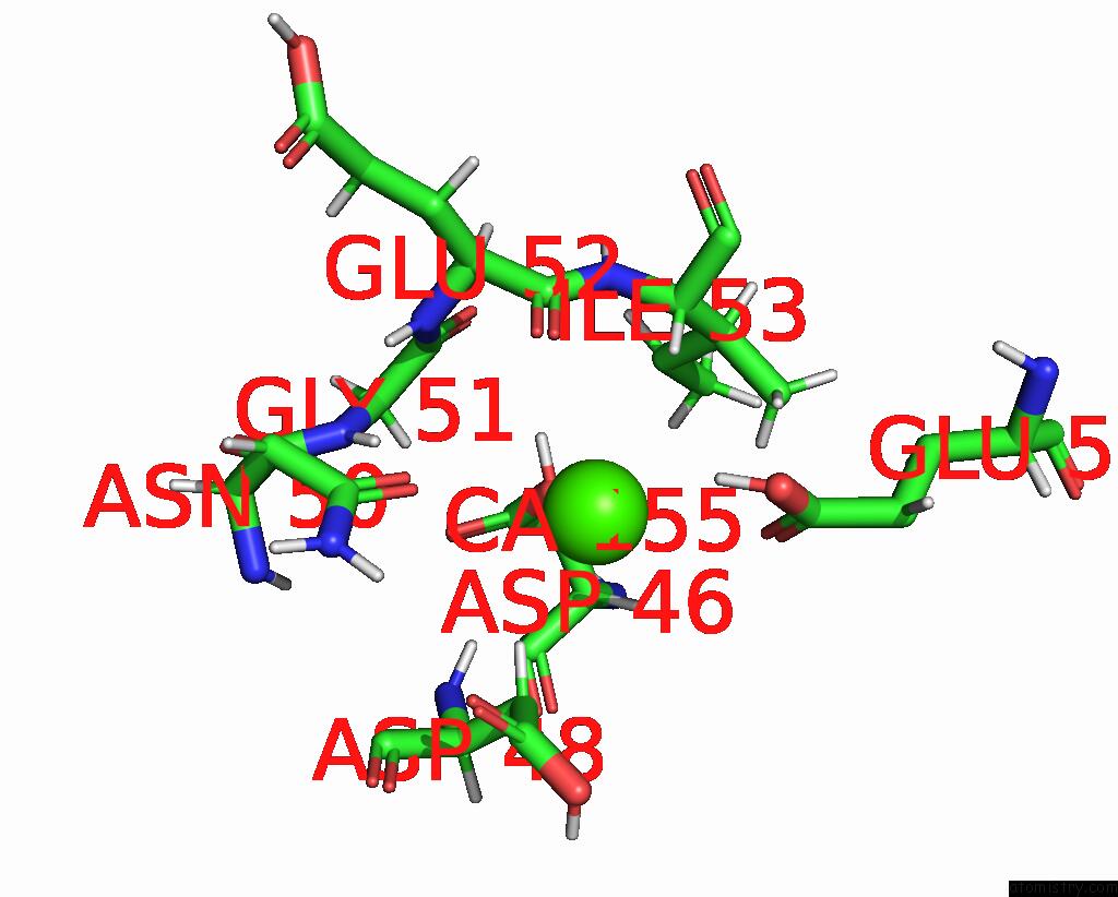
Mono view
Stereo pair view
Mono view
Stereo pair view
A full contact list of Calcium with other atoms in the Ca binding
site number 2 of uc(Nmr) Derived Solution Structure of An Ef-Hand Calcium Binding Protein From Entamoeba Histolytica within 5.0Å range:
|
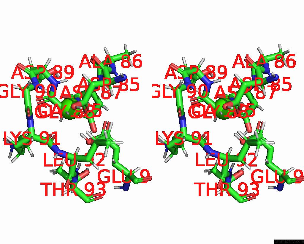
Calcium binding site 3 out of 4 in 2jnx
Go back to
Calcium binding site 3 out
of 4 in the uc(Nmr) Derived Solution Structure of An Ef-Hand Calcium Binding Protein From Entamoeba Histolytica

Mono view
Stereo pair view
Mono view
Stereo pair view
A full contact list of Calcium with other atoms in the Ca binding
site number 3 of uc(Nmr) Derived Solution Structure of An Ef-Hand Calcium Binding Protein From Entamoeba Histolytica within 5.0Å range:
|
Calcium binding site 4 out of 4 in 2jnx
Go back to
Calcium binding site 4 out
of 4 in the uc(Nmr) Derived Solution Structure of An Ef-Hand Calcium Binding Protein From Entamoeba Histolytica
Mono view
Stereo pair view
Mono view
Stereo pair view
A full contact list of Calcium with other atoms in the Ca binding
site number 4 of uc(Nmr) Derived Solution Structure of An Ef-Hand Calcium Binding Protein From Entamoeba Histolytica within 5.0Å range:
|
Reference:
S.M.Mustafi,
R.B.Mutalik.
Structure, Dynamics, and Physiological Properties of A Calcium Binding Protein From Entamoeba Histolytica (EHCABP2) To Be Published.
Page generated: Fri Jul 12 13:48:33 2024
Last articles
Zn in 9JPJZn in 9JP7
Zn in 9JPK
Zn in 9JPL
Zn in 9GN6
Zn in 9GN7
Zn in 9GKU
Zn in 9GKW
Zn in 9GKX
Zn in 9GL0