Calcium »
PDB 2jq6-2ksp »
2jxy »
Calcium in PDB 2jxy: Solution Structure of the Hemopexin-Like Domain of MMP12
Enzymatic activity of Solution Structure of the Hemopexin-Like Domain of MMP12
All present enzymatic activity of Solution Structure of the Hemopexin-Like Domain of MMP12:
3.4.24.65;
3.4.24.65;
Calcium Binding Sites:
The binding sites of Calcium atom in the Solution Structure of the Hemopexin-Like Domain of MMP12
(pdb code 2jxy). This binding sites where shown within
5.0 Angstroms radius around Calcium atom.
In total only one binding site of Calcium was determined in the Solution Structure of the Hemopexin-Like Domain of MMP12, PDB code: 2jxy:
In total only one binding site of Calcium was determined in the Solution Structure of the Hemopexin-Like Domain of MMP12, PDB code: 2jxy:
Calcium binding site 1 out of 1 in 2jxy
Go back to
Calcium binding site 1 out
of 1 in the Solution Structure of the Hemopexin-Like Domain of MMP12
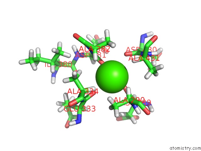
Mono view
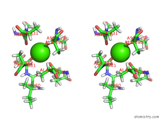
Stereo pair view
Mono view
Stereo pair view
A full contact list of Calcium with other atoms in the Ca binding
site number 1 of Solution Structure of the Hemopexin-Like Domain of MMP12 within 5.0Å range:
|
Reference:
I.Bertini,
V.Calderone,
M.Fragai,
R.Jaiswal,
C.Luchinat,
M.Melikian,
E.Mylonas,
D.I.Svergun.
Evidence of Reciprocal Reorientation of the Catalytic and Hemopexin-Like Domains of Full-Length Mmp-12 J.Am.Chem.Soc. V. 130 7011 2008.
ISSN: ISSN 0002-7863
PubMed: 18465858
DOI: 10.1021/JA710491Y
Page generated: Fri Jul 12 13:52:17 2024
ISSN: ISSN 0002-7863
PubMed: 18465858
DOI: 10.1021/JA710491Y
Last articles
Zn in 9J0NZn in 9J0O
Zn in 9J0P
Zn in 9FJX
Zn in 9EKB
Zn in 9C0F
Zn in 9CAH
Zn in 9CH0
Zn in 9CH3
Zn in 9CH1