Atomistry » Calcium » PDB 2jq6-2ksp » 2kfg
Atomistry »
  Calcium »
    PDB 2jq6-2ksp »
      2kfg »

Calcium in PDB 2kfg: Structure of the C-Terminal Domain of EHD1 in Complex with Fnyestdpftak

Calcium Binding Sites:

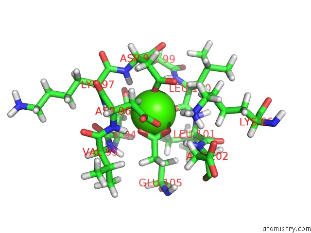
The binding sites of Calcium atom in the Structure of the C-Terminal Domain of EHD1 in Complex with Fnyestdpftak (pdb code 2kfg). This binding sites where shown within 5.0 Angstroms radius around Calcium atom.
In total only one binding site of Calcium was determined in the Structure of the C-Terminal Domain of EHD1 in Complex with Fnyestdpftak, PDB code: 2kfg:

Calcium binding site 1 out of 1 in 2kfg

Go back to Calcium Binding Sites List in 2kfg
Calcium binding site 1 out of 1 in the Structure of the C-Terminal Domain of EHD1 in Complex with Fnyestdpftak


Mono view


Stereo pair view

A full contact list of Calcium with other atoms in the Ca binding site number 1 of Structure of the C-Terminal Domain of EHD1 in Complex with Fnyestdpftak within 5.0Å range:
probe atom residue distance (Å) B Occ
A:Ca141

b:0.5
occ:1.00
OD1 A:ASP98 1.7 0.6 1.0
OD1 A:ASP94 1.8 0.3 1.0
OE2 A:GLU105 1.9 0.4 1.0
OE1 A:GLU105 1.9 0.3 1.0
O A:LEU100 2.0 0.4 1.0
CD A:GLU105 2.2 0.3 1.0
HB2 A:ASP96 2.6 1.6 1.0
CG A:ASP98 2.9 0.6 1.0
CG A:ASP94 3.0 0.2 1.0
H A:ASP98 3.0 0.5 1.0
C A:LEU100 3.1 0.3 1.0
H A:LEU100 3.1 0.4 1.0
H A:ASP96 3.1 0.7 1.0
HA A:LEU101 3.3 0.3 1.0
HA A:ASP94 3.3 0.2 1.0
OD2 A:ASP96 3.4 1.1 1.0
OD2 A:ASP98 3.5 0.7 1.0
H A:ASP102 3.6 0.3 1.0
HD21 A:LEU101 3.6 1.0 1.0
CB A:ASP96 3.6 1.0 1.0
HB2 A:LEU100 3.6 1.0 1.0
CG A:GLU105 3.6 0.2 1.0
H A:LYS97 3.6 0.6 1.0
N A:LEU100 3.7 0.3 1.0
OD2 A:ASP94 3.7 0.3 1.0
CA A:LEU100 3.9 0.3 1.0
N A:ASP98 3.9 0.6 1.0
N A:ASP96 3.9 0.7 1.0
HB3 A:GLU105 4.0 0.2 1.0
CG A:ASP96 4.0 0.8 1.0
HZ1 A:LYS66 4.0 1.5 1.0
N A:LEU101 4.0 0.3 1.0
CA A:ASP94 4.0 0.2 1.0
CB A:ASP94 4.1 0.3 1.0
HG2 A:GLU105 4.1 0.3 1.0
CB A:ASP98 4.1 0.7 1.0
CA A:LEU101 4.1 0.3 1.0
N A:LYS97 4.1 0.7 1.0
H A:GLY99 4.2 0.4 1.0
H A:VAL95 4.2 0.6 1.0
CA A:ASP96 4.2 0.8 1.0
HG3 A:GLU105 4.2 0.3 1.0
C A:ASP94 4.2 0.3 1.0
CB A:LEU100 4.2 0.5 1.0
CB A:GLU105 4.3 0.2 1.0
HB2 A:GLU105 4.3 0.2 1.0
HD3 A:LYS66 4.3 0.8 1.0
N A:VAL95 4.4 0.5 1.0
HB3 A:ASP96 4.4 1.5 1.0
HB3 A:ASP98 4.4 0.8 1.0
CA A:ASP98 4.4 0.6 1.0
N A:ASP102 4.4 0.3 1.0
N A:GLY99 4.4 0.4 1.0
HB3 A:LEU100 4.6 1.0 1.0
C A:ASP96 4.6 0.8 1.0
HB2 A:ASP94 4.6 0.4 1.0
C A:ASP98 4.6 0.5 1.0
CD2 A:LEU101 4.6 0.3 1.0
HB2 A:ASP102 4.6 1.0 1.0
HA A:LYS97 4.7 0.7 1.0
C A:LEU101 4.8 0.3 1.0
C A:GLY99 4.8 0.3 1.0
O A:ASP94 4.9 0.5 1.0
CA A:LYS97 4.9 0.7 1.0
HA A:LEU100 4.9 0.4 1.0
HB3 A:ASP94 4.9 0.4 1.0
C A:LYS97 4.9 0.7 1.0
H A:LEU101 4.9 0.3 1.0
HD22 A:LEU101 5.0 1.1 1.0
C A:VAL95 5.0 0.7 1.0
HB2 A:ASP98 5.0 0.7 1.0

Reference:

F.Kieken, M.Jovic, M.Tonelli, N.Naslavsky, S.Caplan, P.L.Sorgen. Structural Insight Into the Interaction of Proteins Containing Npf, Dpf, and Gpf Motifs with the C-Terminal Eh-Domain of EHD1. Protein Sci. V. 18 2471 2009.
ISSN: ISSN 0961-8368
PubMed: 19798736
DOI: 10.1002/PRO.258
Page generated: Sat Dec 12 03:44:04 2020

Last articles

Zn in 8WB0
Zn in 8WAX
Zn in 8WAU
Zn in 8WAZ
Zn in 8WAY
Zn in 8WAV
Zn in 8WAW
Zn in 8WAT
Zn in 8W7M
Zn in 8WD3
© Copyright 2008-2020 by atomistry.com
Home   |    Site Map   |    Copyright   |    Contact us   |    Privacy