Calcium »
PDB 2jq6-2ksp »
2knx »
Calcium in PDB 2knx: Solution Structure of Complement Repeat CR17 From Lrp-1
Calcium Binding Sites:
The binding sites of Calcium atom in the Solution Structure of Complement Repeat CR17 From Lrp-1
(pdb code 2knx). This binding sites where shown within
5.0 Angstroms radius around Calcium atom.
In total only one binding site of Calcium was determined in the Solution Structure of Complement Repeat CR17 From Lrp-1, PDB code: 2knx:
In total only one binding site of Calcium was determined in the Solution Structure of Complement Repeat CR17 From Lrp-1, PDB code: 2knx:
Calcium binding site 1 out of 1 in 2knx
Go back to
Calcium binding site 1 out
of 1 in the Solution Structure of Complement Repeat CR17 From Lrp-1
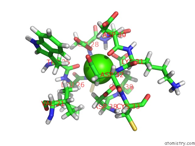
Mono view
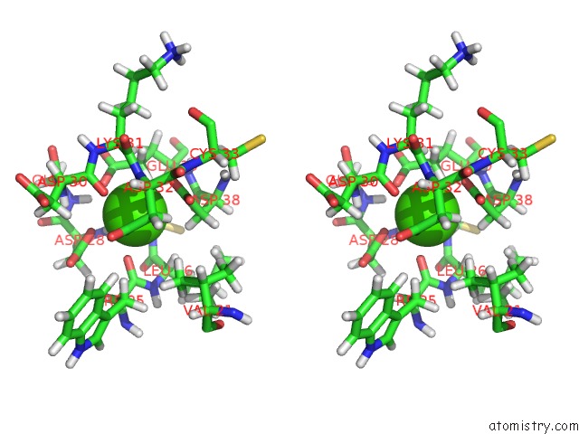
Stereo pair view
Mono view
Stereo pair view
A full contact list of Calcium with other atoms in the Ca binding
site number 1 of Solution Structure of Complement Repeat CR17 From Lrp-1 within 5.0Å range:
|
Reference:
M.Guttman,
J.H.Prieto,
T.M.Handel,
P.J.Domaille,
E.A.Komives.
Structure of the Minimal Interface Between Apoe and Lrp. J.Mol.Biol. V. 398 306 2010.
ISSN: ISSN 0022-2836
PubMed: 20303980
DOI: 10.1016/J.JMB.2010.03.022
Page generated: Fri Jul 12 13:59:23 2024
ISSN: ISSN 0022-2836
PubMed: 20303980
DOI: 10.1016/J.JMB.2010.03.022
Last articles
Zn in 9J0NZn in 9J0O
Zn in 9J0P
Zn in 9FJX
Zn in 9EKB
Zn in 9C0F
Zn in 9CAH
Zn in 9CH0
Zn in 9CH3
Zn in 9CH1