Calcium »
PDB 2m0k-2n8y »
2msb »
Calcium in PDB 2msb: Structure of A C-Type Mannose-Binding Protein Complexed with An Oligosaccharide
Protein crystallography data
The structure of Structure of A C-Type Mannose-Binding Protein Complexed with An Oligosaccharide, PDB code: 2msb
was solved by
W.I.Weis,
K.Drickamer,
W.A.Hendrickson,
with X-Ray Crystallography technique. A brief refinement statistics is given in the table below:
| Resolution Low / High (Å) | 10.00 / 1.70 |
| Space group | P 1 21 1 |
| Cell size a, b, c (Å), α, β, γ (°) | 34.810, 85.160, 39.550, 90.00, 97.41, 90.00 |
| R / Rfree (%) | 17.4 / n/a |
Calcium Binding Sites:
The binding sites of Calcium atom in the Structure of A C-Type Mannose-Binding Protein Complexed with An Oligosaccharide
(pdb code 2msb). This binding sites where shown within
5.0 Angstroms radius around Calcium atom.
In total 6 binding sites of Calcium where determined in the Structure of A C-Type Mannose-Binding Protein Complexed with An Oligosaccharide, PDB code: 2msb:
Jump to Calcium binding site number: 1; 2; 3; 4; 5; 6;
In total 6 binding sites of Calcium where determined in the Structure of A C-Type Mannose-Binding Protein Complexed with An Oligosaccharide, PDB code: 2msb:
Jump to Calcium binding site number: 1; 2; 3; 4; 5; 6;
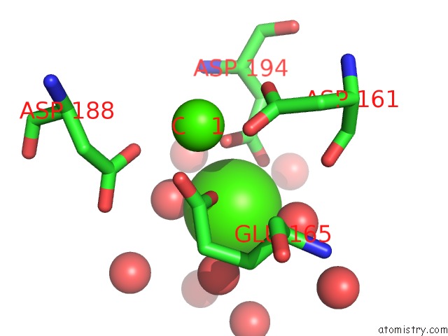
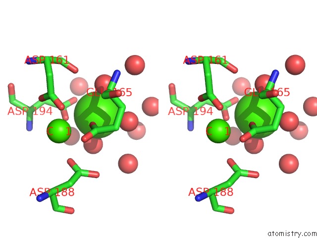
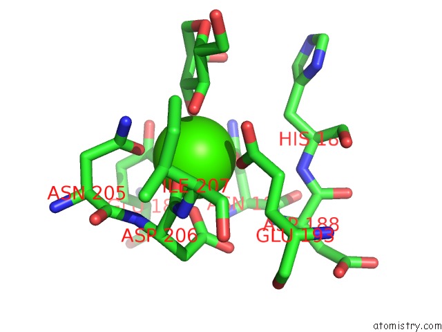
Calcium binding site 1 out of 6 in 2msb
Go back to
Calcium binding site 1 out
of 6 in the Structure of A C-Type Mannose-Binding Protein Complexed with An Oligosaccharide
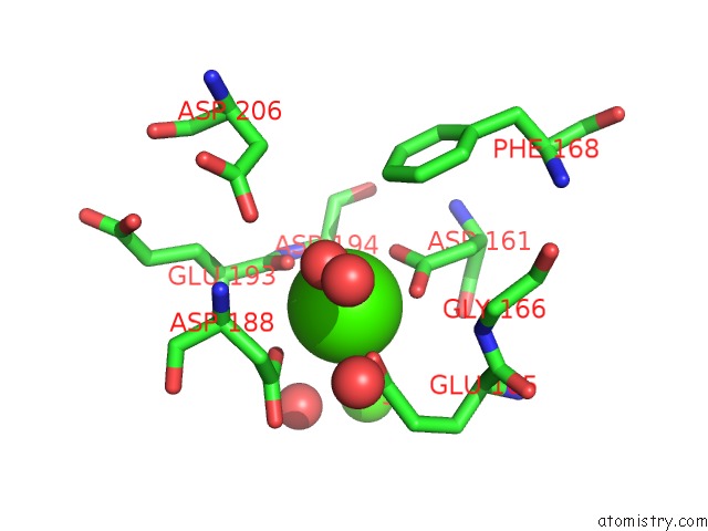
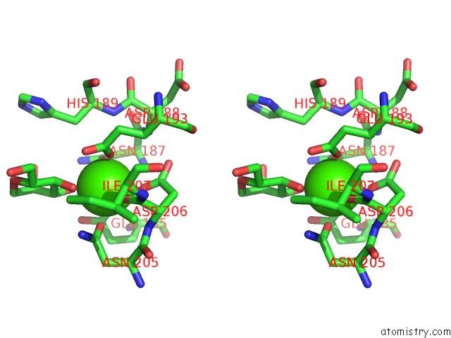
Mono view
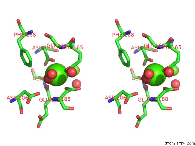
Stereo pair view
Mono view
Stereo pair view
A full contact list of Calcium with other atoms in the Ca binding
site number 1 of Structure of A C-Type Mannose-Binding Protein Complexed with An Oligosaccharide within 5.0Å range:
|
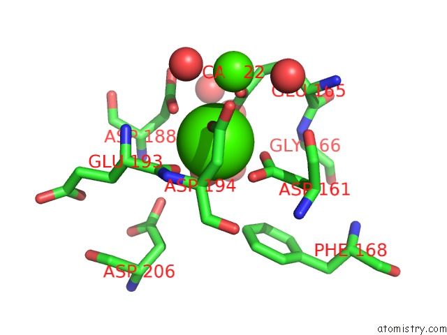
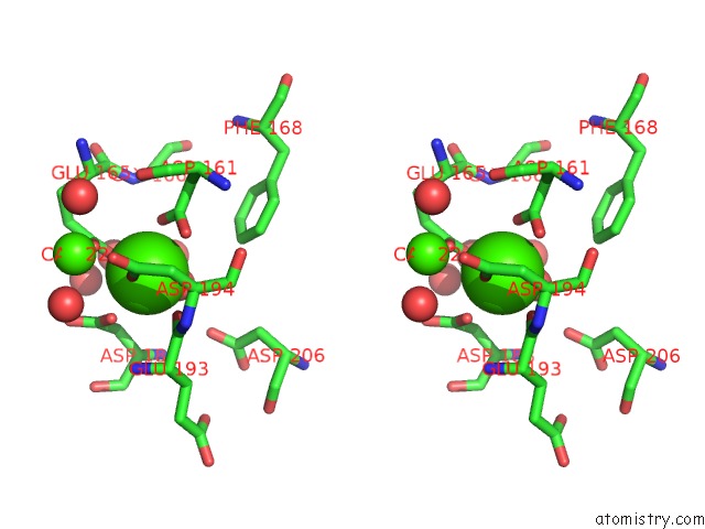
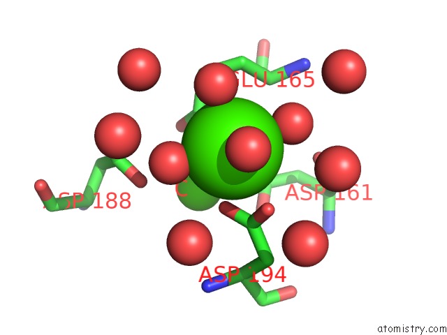
Calcium binding site 2 out of 6 in 2msb
Go back to
Calcium binding site 2 out
of 6 in the Structure of A C-Type Mannose-Binding Protein Complexed with An Oligosaccharide
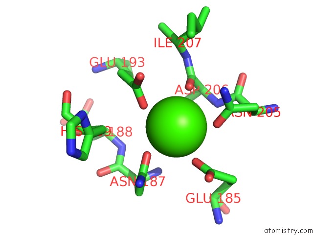
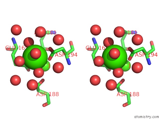
Mono view
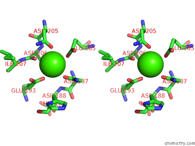
Stereo pair view
Mono view
Stereo pair view
A full contact list of Calcium with other atoms in the Ca binding
site number 2 of Structure of A C-Type Mannose-Binding Protein Complexed with An Oligosaccharide within 5.0Å range:
|
Calcium binding site 3 out of 6 in 2msb
Go back to
Calcium binding site 3 out
of 6 in the Structure of A C-Type Mannose-Binding Protein Complexed with An Oligosaccharide
Mono view
Stereo pair view
Mono view
Stereo pair view
A full contact list of Calcium with other atoms in the Ca binding
site number 3 of Structure of A C-Type Mannose-Binding Protein Complexed with An Oligosaccharide within 5.0Å range:
|
Calcium binding site 4 out of 6 in 2msb
Go back to
Calcium binding site 4 out
of 6 in the Structure of A C-Type Mannose-Binding Protein Complexed with An Oligosaccharide
Mono view
Stereo pair view
Mono view
Stereo pair view
A full contact list of Calcium with other atoms in the Ca binding
site number 4 of Structure of A C-Type Mannose-Binding Protein Complexed with An Oligosaccharide within 5.0Å range:
|
Calcium binding site 5 out of 6 in 2msb
Go back to
Calcium binding site 5 out
of 6 in the Structure of A C-Type Mannose-Binding Protein Complexed with An Oligosaccharide
Mono view
Stereo pair view
Mono view
Stereo pair view
A full contact list of Calcium with other atoms in the Ca binding
site number 5 of Structure of A C-Type Mannose-Binding Protein Complexed with An Oligosaccharide within 5.0Å range:
|
Calcium binding site 6 out of 6 in 2msb
Go back to
Calcium binding site 6 out
of 6 in the Structure of A C-Type Mannose-Binding Protein Complexed with An Oligosaccharide
Mono view
Stereo pair view
Mono view
Stereo pair view
A full contact list of Calcium with other atoms in the Ca binding
site number 6 of Structure of A C-Type Mannose-Binding Protein Complexed with An Oligosaccharide within 5.0Å range:
|
Reference:
W.I.Weis,
K.Drickamer,
W.A.Hendrickson.
Structure of A C-Type Mannose-Binding Protein Complexed with An Oligosaccharide. Nature V. 360 127 1992.
ISSN: ISSN 0028-0836
PubMed: 1436090
DOI: 10.1038/360127A0
Page generated: Fri Jul 12 14:22:07 2024
ISSN: ISSN 0028-0836
PubMed: 1436090
DOI: 10.1038/360127A0
Last articles
Zn in 9MJ5Zn in 9HNW
Zn in 9G0L
Zn in 9FNE
Zn in 9DZN
Zn in 9E0I
Zn in 9D32
Zn in 9DAK
Zn in 8ZXC
Zn in 8ZUF