Calcium »
PDB 2nce-2o86 »
2np0 »
Calcium in PDB 2np0: Crystal Structure of the Botulinum Neurotoxin Type B Complexed with Synaptotagamin-II Ectodomain
Enzymatic activity of Crystal Structure of the Botulinum Neurotoxin Type B Complexed with Synaptotagamin-II Ectodomain
All present enzymatic activity of Crystal Structure of the Botulinum Neurotoxin Type B Complexed with Synaptotagamin-II Ectodomain:
3.4.24.69;
3.4.24.69;
Protein crystallography data
The structure of Crystal Structure of the Botulinum Neurotoxin Type B Complexed with Synaptotagamin-II Ectodomain, PDB code: 2np0
was solved by
Q.Chai,
J.W.Arndt,
R.C.Stevens,
with X-Ray Crystallography technique. A brief refinement statistics is given in the table below:
| Resolution Low / High (Å) | 40.00 / 2.62 |
| Space group | C 2 2 21 |
| Cell size a, b, c (Å), α, β, γ (°) | 170.415, 351.971, 101.968, 90.00, 90.00, 90.00 |
| R / Rfree (%) | 18.6 / 22.7 |
Other elements in 2np0:
The structure of Crystal Structure of the Botulinum Neurotoxin Type B Complexed with Synaptotagamin-II Ectodomain also contains other interesting chemical elements:
| Chlorine | (Cl) | 1 atom |
| Zinc | (Zn) | 1 atom |
Calcium Binding Sites:
The binding sites of Calcium atom in the Crystal Structure of the Botulinum Neurotoxin Type B Complexed with Synaptotagamin-II Ectodomain
(pdb code 2np0). This binding sites where shown within
5.0 Angstroms radius around Calcium atom.
In total 3 binding sites of Calcium where determined in the Crystal Structure of the Botulinum Neurotoxin Type B Complexed with Synaptotagamin-II Ectodomain, PDB code: 2np0:
Jump to Calcium binding site number: 1; 2; 3;
In total 3 binding sites of Calcium where determined in the Crystal Structure of the Botulinum Neurotoxin Type B Complexed with Synaptotagamin-II Ectodomain, PDB code: 2np0:
Jump to Calcium binding site number: 1; 2; 3;
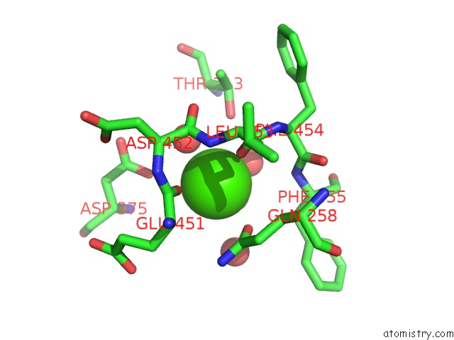
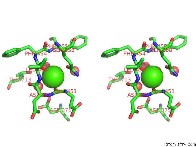
Calcium binding site 1 out of 3 in 2np0
Go back to
Calcium binding site 1 out
of 3 in the Crystal Structure of the Botulinum Neurotoxin Type B Complexed with Synaptotagamin-II Ectodomain
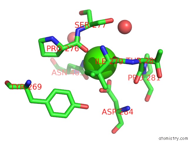
Mono view
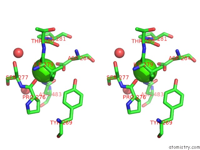
Stereo pair view
Mono view
Stereo pair view
A full contact list of Calcium with other atoms in the Ca binding
site number 1 of Crystal Structure of the Botulinum Neurotoxin Type B Complexed with Synaptotagamin-II Ectodomain within 5.0Å range:
|
Calcium binding site 2 out of 3 in 2np0
Go back to
Calcium binding site 2 out
of 3 in the Crystal Structure of the Botulinum Neurotoxin Type B Complexed with Synaptotagamin-II Ectodomain
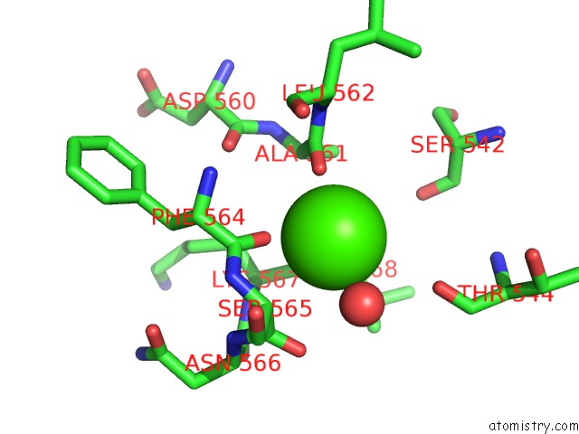
Mono view
Stereo pair view
Mono view
Stereo pair view
A full contact list of Calcium with other atoms in the Ca binding
site number 2 of Crystal Structure of the Botulinum Neurotoxin Type B Complexed with Synaptotagamin-II Ectodomain within 5.0Å range:
|
Calcium binding site 3 out of 3 in 2np0
Go back to
Calcium binding site 3 out
of 3 in the Crystal Structure of the Botulinum Neurotoxin Type B Complexed with Synaptotagamin-II Ectodomain
Mono view
Stereo pair view
Mono view
Stereo pair view
A full contact list of Calcium with other atoms in the Ca binding
site number 3 of Crystal Structure of the Botulinum Neurotoxin Type B Complexed with Synaptotagamin-II Ectodomain within 5.0Å range:
|
Reference:
Q.Chai,
J.W.Arndt,
M.Dong,
W.H.Tepp,
E.A.Johnson,
E.R.Chapman,
R.C.Stevens.
Structural Basis of Cell Surface Receptor Recognition By Botulinum Neurotoxin B. Nature V. 444 1096 2006.
ISSN: ISSN 0028-0836
PubMed: 17167418
DOI: 10.1038/NATURE05411
Page generated: Fri Jul 12 14:27:48 2024
ISSN: ISSN 0028-0836
PubMed: 17167418
DOI: 10.1038/NATURE05411
Last articles
Zn in 9J0NZn in 9J0O
Zn in 9J0P
Zn in 9FJX
Zn in 9EKB
Zn in 9C0F
Zn in 9CAH
Zn in 9CH0
Zn in 9CH3
Zn in 9CH1