Calcium »
PDB 2nce-2o86 »
2nz9 »
Calcium in PDB 2nz9: Crystal Structure of Botulinum Neurotoxin Type A Complexed with Monoclonal Antibody AR2
Enzymatic activity of Crystal Structure of Botulinum Neurotoxin Type A Complexed with Monoclonal Antibody AR2
All present enzymatic activity of Crystal Structure of Botulinum Neurotoxin Type A Complexed with Monoclonal Antibody AR2:
3.4.24.69;
3.4.24.69;
Protein crystallography data
The structure of Crystal Structure of Botulinum Neurotoxin Type A Complexed with Monoclonal Antibody AR2, PDB code: 2nz9
was solved by
R.C.Stevens,
J.W.Arndt,
with X-Ray Crystallography technique. A brief refinement statistics is given in the table below:
| Resolution Low / High (Å) | 40.00 / 3.79 |
| Space group | P 1 21 1 |
| Cell size a, b, c (Å), α, β, γ (°) | 97.965, 197.596, 146.105, 90.00, 91.18, 90.00 |
| R / Rfree (%) | 22.4 / 27.8 |
Other elements in 2nz9:
The structure of Crystal Structure of Botulinum Neurotoxin Type A Complexed with Monoclonal Antibody AR2 also contains other interesting chemical elements:
| Zinc | (Zn) | 2 atoms |
Calcium Binding Sites:
The binding sites of Calcium atom in the Crystal Structure of Botulinum Neurotoxin Type A Complexed with Monoclonal Antibody AR2
(pdb code 2nz9). This binding sites where shown within
5.0 Angstroms radius around Calcium atom.
In total 2 binding sites of Calcium where determined in the Crystal Structure of Botulinum Neurotoxin Type A Complexed with Monoclonal Antibody AR2, PDB code: 2nz9:
Jump to Calcium binding site number: 1; 2;
In total 2 binding sites of Calcium where determined in the Crystal Structure of Botulinum Neurotoxin Type A Complexed with Monoclonal Antibody AR2, PDB code: 2nz9:
Jump to Calcium binding site number: 1; 2;
Calcium binding site 1 out of 2 in 2nz9
Go back to
Calcium binding site 1 out
of 2 in the Crystal Structure of Botulinum Neurotoxin Type A Complexed with Monoclonal Antibody AR2
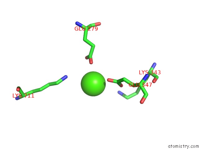
Mono view
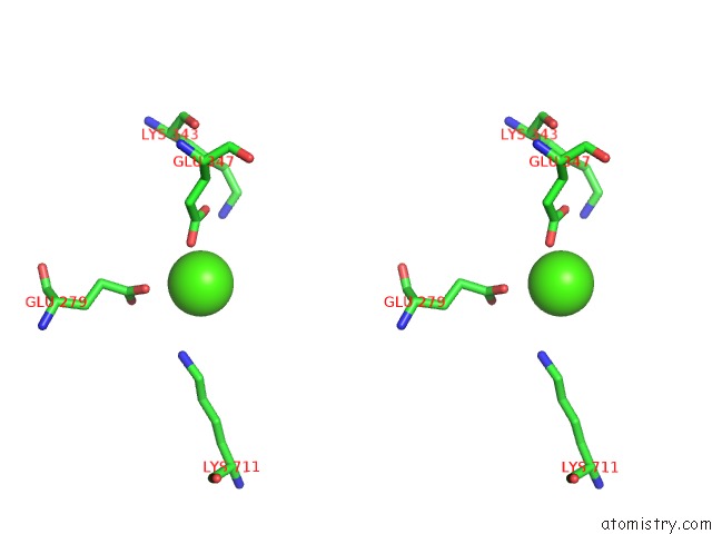
Stereo pair view
Mono view
Stereo pair view
A full contact list of Calcium with other atoms in the Ca binding
site number 1 of Crystal Structure of Botulinum Neurotoxin Type A Complexed with Monoclonal Antibody AR2 within 5.0Å range:
|
Calcium binding site 2 out of 2 in 2nz9
Go back to
Calcium binding site 2 out
of 2 in the Crystal Structure of Botulinum Neurotoxin Type A Complexed with Monoclonal Antibody AR2
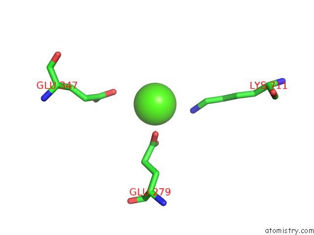
Mono view
Stereo pair view
Mono view
Stereo pair view
A full contact list of Calcium with other atoms in the Ca binding
site number 2 of Crystal Structure of Botulinum Neurotoxin Type A Complexed with Monoclonal Antibody AR2 within 5.0Å range:
|
Reference:
C.Garcia-Rodriguez,
R.Levy,
J.W.Arndt,
C.M.Forsyth,
A.Razai,
J.Lou,
I.Geren,
R.C.Stevens,
J.D.Marks.
Molecular Evolution of Antibody Cross-Reactivity For Two Subtypes of Type A Botulinum Neurotoxin. Nat.Biotechnol. V. 25 107 2007.
ISSN: ISSN 1087-0156
PubMed: 17173035
DOI: 10.1038/NBT1269
Page generated: Tue Jul 8 07:14:05 2025
ISSN: ISSN 1087-0156
PubMed: 17173035
DOI: 10.1038/NBT1269
Last articles
Cl in 5RT6Cl in 5RTH
Cl in 5RTI
Cl in 5ROQ
Cl in 5RSL
Cl in 5ROW
Cl in 5RMJ
Cl in 5ROP
Cl in 5RML
Cl in 5RKR