Calcium »
PDB 2o8o-2ovu »
2oot »
Calcium in PDB 2oot: A High Resolution Structure of Ligand-Free Human Glutamate Carboxypeptidase II
Enzymatic activity of A High Resolution Structure of Ligand-Free Human Glutamate Carboxypeptidase II
All present enzymatic activity of A High Resolution Structure of Ligand-Free Human Glutamate Carboxypeptidase II:
3.4.17.21;
3.4.17.21;
Protein crystallography data
The structure of A High Resolution Structure of Ligand-Free Human Glutamate Carboxypeptidase II, PDB code: 2oot
was solved by
C.Barinka,
J.Lubkowski,
with X-Ray Crystallography technique. A brief refinement statistics is given in the table below:
| Resolution Low / High (Å) | 30.00 / 1.64 |
| Space group | I 2 2 2 |
| Cell size a, b, c (Å), α, β, γ (°) | 101.759, 130.128, 158.874, 90.00, 90.00, 90.00 |
| R / Rfree (%) | 20.6 / 22.8 |
Other elements in 2oot:
The structure of A High Resolution Structure of Ligand-Free Human Glutamate Carboxypeptidase II also contains other interesting chemical elements:
| Chlorine | (Cl) | 1 atom |
| Zinc | (Zn) | 2 atoms |
Calcium Binding Sites:
The binding sites of Calcium atom in the A High Resolution Structure of Ligand-Free Human Glutamate Carboxypeptidase II
(pdb code 2oot). This binding sites where shown within
5.0 Angstroms radius around Calcium atom.
In total only one binding site of Calcium was determined in the A High Resolution Structure of Ligand-Free Human Glutamate Carboxypeptidase II, PDB code: 2oot:
In total only one binding site of Calcium was determined in the A High Resolution Structure of Ligand-Free Human Glutamate Carboxypeptidase II, PDB code: 2oot:
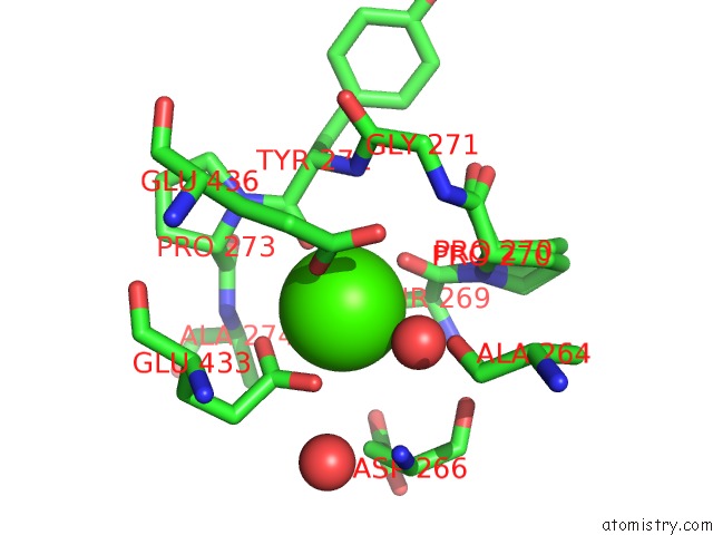
Calcium binding site 1 out of 1 in 2oot
Go back to
Calcium binding site 1 out
of 1 in the A High Resolution Structure of Ligand-Free Human Glutamate Carboxypeptidase II
Mono view

Stereo pair view
Mono view
Stereo pair view
A full contact list of Calcium with other atoms in the Ca binding
site number 1 of A High Resolution Structure of Ligand-Free Human Glutamate Carboxypeptidase II within 5.0Å range:
|
Reference:
C.Barinka,
J.Starkova,
J.Konvalinka,
J.Lubkowski.
A High-Resolution Structure of Ligand-Free Human Glutamate Carboxypeptidase II. Acta Crystallogr.,Sect.F V. 63 150 2007.
ISSN: ESSN 1744-3091
PubMed: 17329803
DOI: 10.1107/S174430910700379X
Page generated: Tue Jul 8 07:21:36 2025
ISSN: ESSN 1744-3091
PubMed: 17329803
DOI: 10.1107/S174430910700379X
Last articles
Ca in 7G5UCa in 7G5V
Ca in 7G5W
Ca in 7G5T
Ca in 7G5R
Ca in 7G5S
Ca in 7G5Q
Ca in 7G5N
Ca in 7G5P
Ca in 7G5O