Calcium »
PDB 2o8o-2ovu »
2os9 »
Calcium in PDB 2os9: Crystal Structure of the Trimeric Neck and Carbohydrate Recognition Domain of Human Surfactant Protein D in Complex with Myoinositol
Protein crystallography data
The structure of Crystal Structure of the Trimeric Neck and Carbohydrate Recognition Domain of Human Surfactant Protein D in Complex with Myoinositol, PDB code: 2os9
was solved by
J.F.Head,
with X-Ray Crystallography technique. A brief refinement statistics is given in the table below:
| Resolution Low / High (Å) | 27.87 / 1.70 |
| Space group | P 1 21 1 |
| Cell size a, b, c (Å), α, β, γ (°) | 55.564, 108.692, 55.774, 90.00, 91.49, 90.00 |
| R / Rfree (%) | 19.5 / 22.3 |
Calcium Binding Sites:
The binding sites of Calcium atom in the Crystal Structure of the Trimeric Neck and Carbohydrate Recognition Domain of Human Surfactant Protein D in Complex with Myoinositol
(pdb code 2os9). This binding sites where shown within
5.0 Angstroms radius around Calcium atom.
In total 9 binding sites of Calcium where determined in the Crystal Structure of the Trimeric Neck and Carbohydrate Recognition Domain of Human Surfactant Protein D in Complex with Myoinositol, PDB code: 2os9:
Jump to Calcium binding site number: 1; 2; 3; 4; 5; 6; 7; 8; 9;
In total 9 binding sites of Calcium where determined in the Crystal Structure of the Trimeric Neck and Carbohydrate Recognition Domain of Human Surfactant Protein D in Complex with Myoinositol, PDB code: 2os9:
Jump to Calcium binding site number: 1; 2; 3; 4; 5; 6; 7; 8; 9;
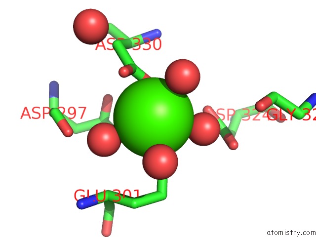
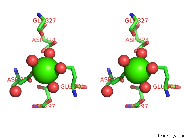
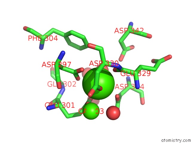
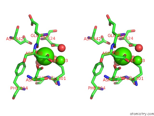
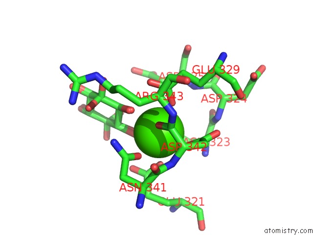
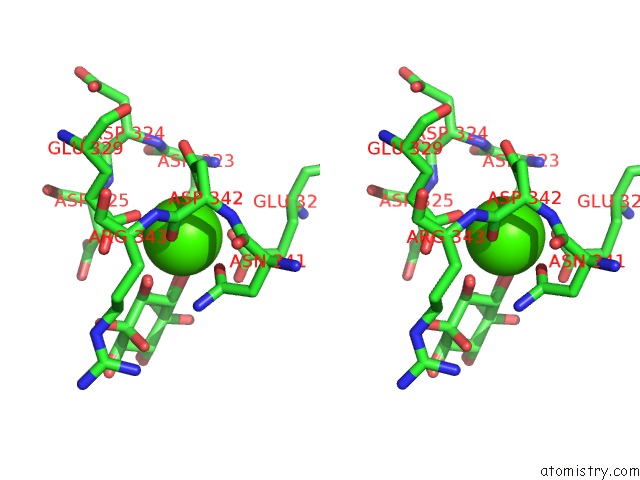
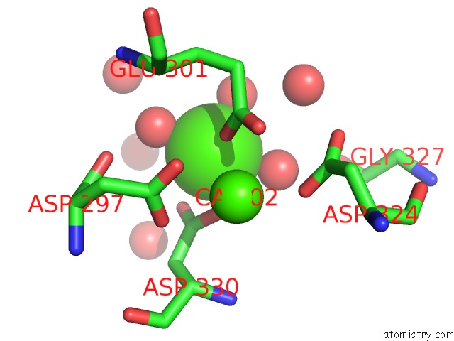
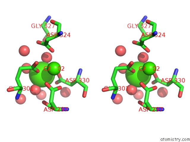
Calcium binding site 1 out of 9 in 2os9
Go back to
Calcium binding site 1 out
of 9 in the Crystal Structure of the Trimeric Neck and Carbohydrate Recognition Domain of Human Surfactant Protein D in Complex with Myoinositol
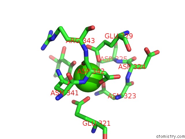
Mono view
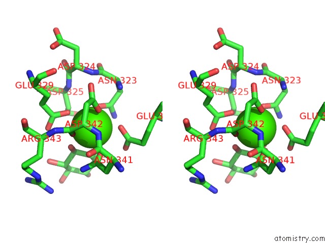
Stereo pair view
Mono view
Stereo pair view
A full contact list of Calcium with other atoms in the Ca binding
site number 1 of Crystal Structure of the Trimeric Neck and Carbohydrate Recognition Domain of Human Surfactant Protein D in Complex with Myoinositol within 5.0Å range:
|
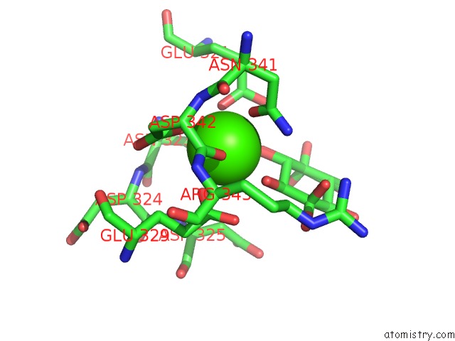
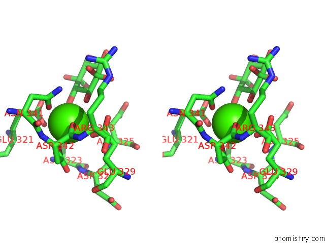
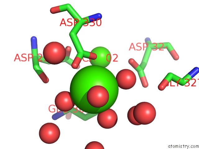
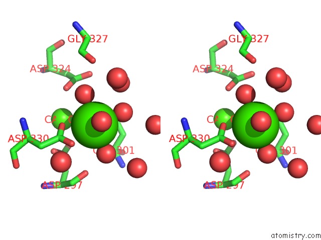
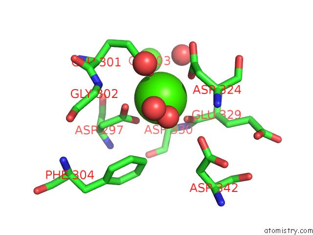
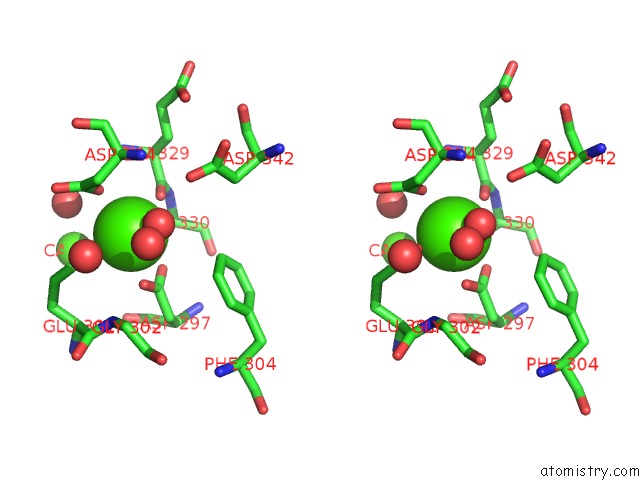
Calcium binding site 2 out of 9 in 2os9
Go back to
Calcium binding site 2 out
of 9 in the Crystal Structure of the Trimeric Neck and Carbohydrate Recognition Domain of Human Surfactant Protein D in Complex with Myoinositol
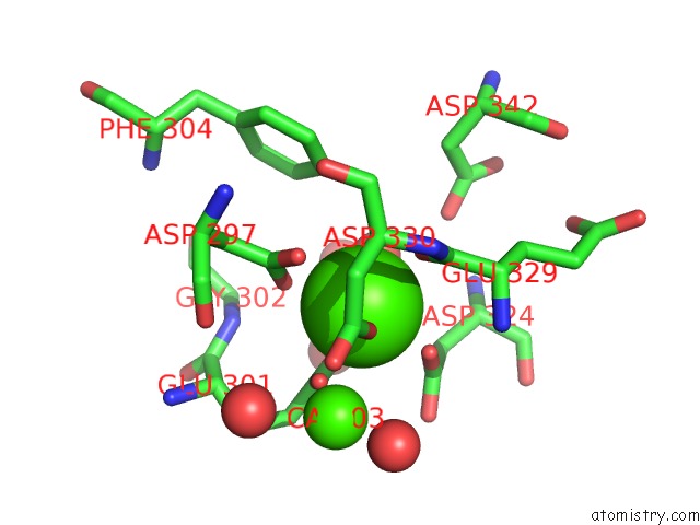
Mono view
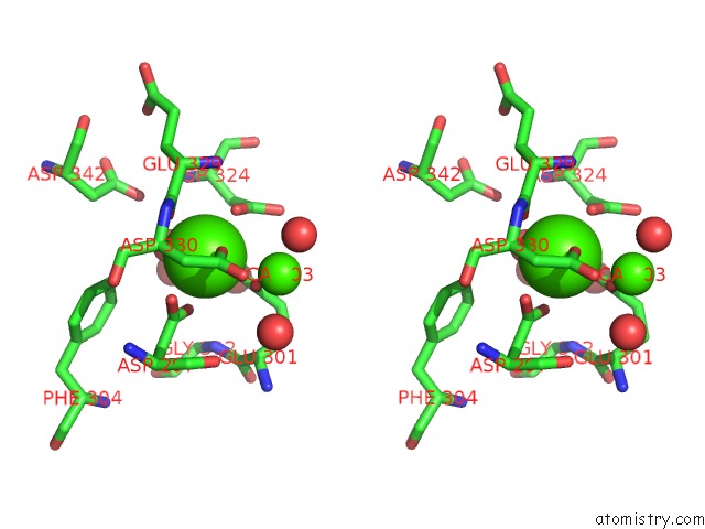
Stereo pair view
Mono view
Stereo pair view
A full contact list of Calcium with other atoms in the Ca binding
site number 2 of Crystal Structure of the Trimeric Neck and Carbohydrate Recognition Domain of Human Surfactant Protein D in Complex with Myoinositol within 5.0Å range:
|
Calcium binding site 3 out of 9 in 2os9
Go back to
Calcium binding site 3 out
of 9 in the Crystal Structure of the Trimeric Neck and Carbohydrate Recognition Domain of Human Surfactant Protein D in Complex with Myoinositol
Mono view
Stereo pair view
Mono view
Stereo pair view
A full contact list of Calcium with other atoms in the Ca binding
site number 3 of Crystal Structure of the Trimeric Neck and Carbohydrate Recognition Domain of Human Surfactant Protein D in Complex with Myoinositol within 5.0Å range:
|
Calcium binding site 4 out of 9 in 2os9
Go back to
Calcium binding site 4 out
of 9 in the Crystal Structure of the Trimeric Neck and Carbohydrate Recognition Domain of Human Surfactant Protein D in Complex with Myoinositol
Mono view
Stereo pair view
Mono view
Stereo pair view
A full contact list of Calcium with other atoms in the Ca binding
site number 4 of Crystal Structure of the Trimeric Neck and Carbohydrate Recognition Domain of Human Surfactant Protein D in Complex with Myoinositol within 5.0Å range:
|
Calcium binding site 5 out of 9 in 2os9
Go back to
Calcium binding site 5 out
of 9 in the Crystal Structure of the Trimeric Neck and Carbohydrate Recognition Domain of Human Surfactant Protein D in Complex with Myoinositol
Mono view
Stereo pair view
Mono view
Stereo pair view
A full contact list of Calcium with other atoms in the Ca binding
site number 5 of Crystal Structure of the Trimeric Neck and Carbohydrate Recognition Domain of Human Surfactant Protein D in Complex with Myoinositol within 5.0Å range:
|
Calcium binding site 6 out of 9 in 2os9
Go back to
Calcium binding site 6 out
of 9 in the Crystal Structure of the Trimeric Neck and Carbohydrate Recognition Domain of Human Surfactant Protein D in Complex with Myoinositol
Mono view
Stereo pair view
Mono view
Stereo pair view
A full contact list of Calcium with other atoms in the Ca binding
site number 6 of Crystal Structure of the Trimeric Neck and Carbohydrate Recognition Domain of Human Surfactant Protein D in Complex with Myoinositol within 5.0Å range:
|
Calcium binding site 7 out of 9 in 2os9
Go back to
Calcium binding site 7 out
of 9 in the Crystal Structure of the Trimeric Neck and Carbohydrate Recognition Domain of Human Surfactant Protein D in Complex with Myoinositol
Mono view
Stereo pair view
Mono view
Stereo pair view
A full contact list of Calcium with other atoms in the Ca binding
site number 7 of Crystal Structure of the Trimeric Neck and Carbohydrate Recognition Domain of Human Surfactant Protein D in Complex with Myoinositol within 5.0Å range:
|
Calcium binding site 8 out of 9 in 2os9
Go back to
Calcium binding site 8 out
of 9 in the Crystal Structure of the Trimeric Neck and Carbohydrate Recognition Domain of Human Surfactant Protein D in Complex with Myoinositol
Mono view
Stereo pair view
Mono view
Stereo pair view
A full contact list of Calcium with other atoms in the Ca binding
site number 8 of Crystal Structure of the Trimeric Neck and Carbohydrate Recognition Domain of Human Surfactant Protein D in Complex with Myoinositol within 5.0Å range:
|
Calcium binding site 9 out of 9 in 2os9
Go back to
Calcium binding site 9 out
of 9 in the Crystal Structure of the Trimeric Neck and Carbohydrate Recognition Domain of Human Surfactant Protein D in Complex with Myoinositol
Mono view
Stereo pair view
Mono view
Stereo pair view
A full contact list of Calcium with other atoms in the Ca binding
site number 9 of Crystal Structure of the Trimeric Neck and Carbohydrate Recognition Domain of Human Surfactant Protein D in Complex with Myoinositol within 5.0Å range:
|
Reference:
E.Crouch,
B.Mcdonald,
K.Smith,
M.Roberts,
T.Mealy,
B.Seaton,
J.Head.
Critical Role of Arg/LYS343 in the Species-Dependent Recognition of Phosphatidylinositol By Pulmonary Surfactant Protein D. Biochemistry V. 46 5160 2007.
ISSN: ISSN 0006-2960
PubMed: 17417879
DOI: 10.1021/BI700037X
Page generated: Fri Jul 12 14:42:21 2024
ISSN: ISSN 0006-2960
PubMed: 17417879
DOI: 10.1021/BI700037X
Last articles
Zn in 9J0NZn in 9J0O
Zn in 9J0P
Zn in 9FJX
Zn in 9EKB
Zn in 9C0F
Zn in 9CAH
Zn in 9CH0
Zn in 9CH3
Zn in 9CH1