Calcium »
PDB 2ovx-2p9k »
2oyi »
Calcium in PDB 2oyi: Crystal Structure of Fragment D of GAMMAD298,301A Fibrinogen with the Peptide Ligand Gly-Pro-Arg-Pro-Amide
Protein crystallography data
The structure of Crystal Structure of Fragment D of GAMMAD298,301A Fibrinogen with the Peptide Ligand Gly-Pro-Arg-Pro-Amide, PDB code: 2oyi
was solved by
M.S.Kostelansky,
O.V.Gorkun,
S.T.Lord,
with X-Ray Crystallography technique. A brief refinement statistics is given in the table below:
| Resolution Low / High (Å) | 17.97 / 2.70 |
| Space group | P 21 21 21 |
| Cell size a, b, c (Å), α, β, γ (°) | 88.988, 94.038, 226.353, 90.00, 90.00, 90.00 |
| R / Rfree (%) | 21.6 / 25.6 |
Calcium Binding Sites:
The binding sites of Calcium atom in the Crystal Structure of Fragment D of GAMMAD298,301A Fibrinogen with the Peptide Ligand Gly-Pro-Arg-Pro-Amide
(pdb code 2oyi). This binding sites where shown within
5.0 Angstroms radius around Calcium atom.
In total 4 binding sites of Calcium where determined in the Crystal Structure of Fragment D of GAMMAD298,301A Fibrinogen with the Peptide Ligand Gly-Pro-Arg-Pro-Amide, PDB code: 2oyi:
Jump to Calcium binding site number: 1; 2; 3; 4;
In total 4 binding sites of Calcium where determined in the Crystal Structure of Fragment D of GAMMAD298,301A Fibrinogen with the Peptide Ligand Gly-Pro-Arg-Pro-Amide, PDB code: 2oyi:
Jump to Calcium binding site number: 1; 2; 3; 4;
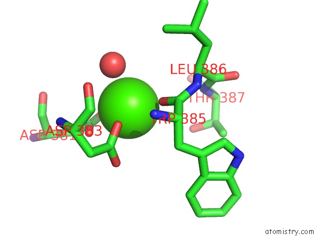
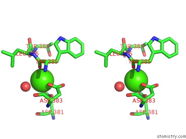
Calcium binding site 1 out of 4 in 2oyi
Go back to
Calcium binding site 1 out
of 4 in the Crystal Structure of Fragment D of GAMMAD298,301A Fibrinogen with the Peptide Ligand Gly-Pro-Arg-Pro-Amide
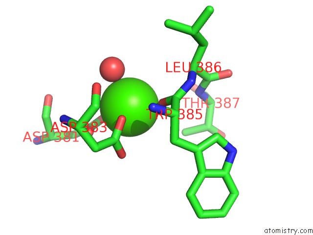
Mono view
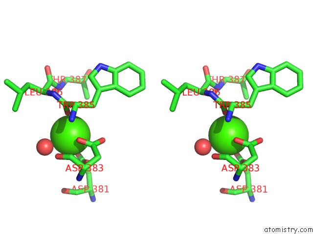
Stereo pair view
Mono view
Stereo pair view
A full contact list of Calcium with other atoms in the Ca binding
site number 1 of Crystal Structure of Fragment D of GAMMAD298,301A Fibrinogen with the Peptide Ligand Gly-Pro-Arg-Pro-Amide within 5.0Å range:
|
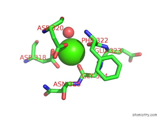
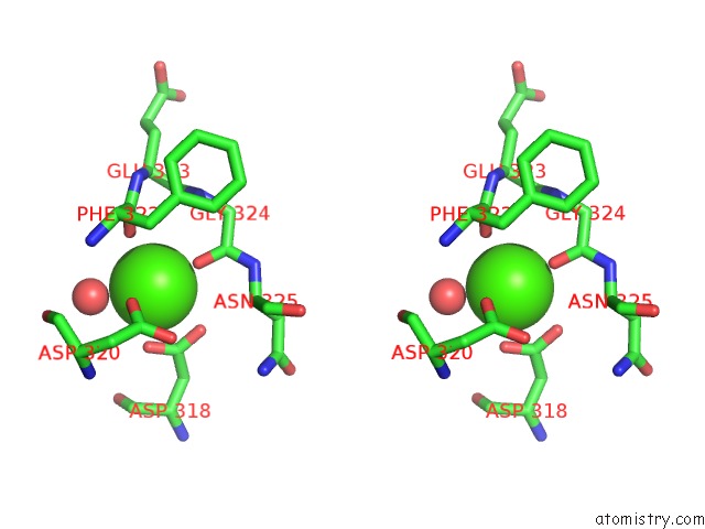
Calcium binding site 2 out of 4 in 2oyi
Go back to
Calcium binding site 2 out
of 4 in the Crystal Structure of Fragment D of GAMMAD298,301A Fibrinogen with the Peptide Ligand Gly-Pro-Arg-Pro-Amide
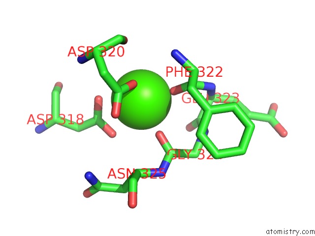
Mono view
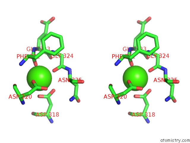
Stereo pair view
Mono view
Stereo pair view
A full contact list of Calcium with other atoms in the Ca binding
site number 2 of Crystal Structure of Fragment D of GAMMAD298,301A Fibrinogen with the Peptide Ligand Gly-Pro-Arg-Pro-Amide within 5.0Å range:
|
Calcium binding site 3 out of 4 in 2oyi
Go back to
Calcium binding site 3 out
of 4 in the Crystal Structure of Fragment D of GAMMAD298,301A Fibrinogen with the Peptide Ligand Gly-Pro-Arg-Pro-Amide
Mono view
Stereo pair view
Mono view
Stereo pair view
A full contact list of Calcium with other atoms in the Ca binding
site number 3 of Crystal Structure of Fragment D of GAMMAD298,301A Fibrinogen with the Peptide Ligand Gly-Pro-Arg-Pro-Amide within 5.0Å range:
|
Calcium binding site 4 out of 4 in 2oyi
Go back to
Calcium binding site 4 out
of 4 in the Crystal Structure of Fragment D of GAMMAD298,301A Fibrinogen with the Peptide Ligand Gly-Pro-Arg-Pro-Amide
Mono view
Stereo pair view
Mono view
Stereo pair view
A full contact list of Calcium with other atoms in the Ca binding
site number 4 of Crystal Structure of Fragment D of GAMMAD298,301A Fibrinogen with the Peptide Ligand Gly-Pro-Arg-Pro-Amide within 5.0Å range:
|
Reference:
M.S.Kostelansky,
K.C.Lounes,
L.F.Ping,
S.K.Dickerson,
O.V.Gorkun,
S.T.Lord.
Probing the GAMMA2 Calcium-Binding Site: Studies with GAMMAD298,301A Fibrinogen Reveal Changes in the GAMMA294-301 Loop That Alter the Integrity of the "A" Polymerization Site. Biochemistry V. 46 5114 2007.
ISSN: ISSN 0006-2960
PubMed: 17411074
DOI: 10.1021/BI602607A
Page generated: Tue Jul 8 07:32:21 2025
ISSN: ISSN 0006-2960
PubMed: 17411074
DOI: 10.1021/BI602607A
Last articles
F in 7LG8F in 7LD3
F in 7LCR
F in 7LCM
F in 7LCO
F in 7LCK
F in 7LCJ
F in 7LCI
F in 7L9Y
F in 7LCD