Calcium »
PDB 2ovx-2p9k »
2p5w »
Calcium in PDB 2p5w: Crystal Structures of High Affinity Human T-Cell Receptors Bound to Pmhc Reveal Native Diagonal Binding Geometry
Protein crystallography data
The structure of Crystal Structures of High Affinity Human T-Cell Receptors Bound to Pmhc Reveal Native Diagonal Binding Geometry, PDB code: 2p5w
was solved by
M.Sami,
P.J.Rizkallah,
S.Dunn,
Y.Li,
R.Moysey,
A.Vuidepot,
E.Baston,
P.Todorov,
P.Molloy,
F.Gao,
J.M.Boulter,
B.K.Jakobsen,
with X-Ray Crystallography technique. A brief refinement statistics is given in the table below:
| Resolution Low / High (Å) | 24.80 / 2.20 |
| Space group | P 1 21 1 |
| Cell size a, b, c (Å), α, β, γ (°) | 74.590, 52.325, 118.932, 90.00, 98.13, 90.00 |
| R / Rfree (%) | 16.5 / 24.5 |
Other elements in 2p5w:
The structure of Crystal Structures of High Affinity Human T-Cell Receptors Bound to Pmhc Reveal Native Diagonal Binding Geometry also contains other interesting chemical elements:
| Magnesium | (Mg) | 1 atom |
Calcium Binding Sites:
The binding sites of Calcium atom in the Crystal Structures of High Affinity Human T-Cell Receptors Bound to Pmhc Reveal Native Diagonal Binding Geometry
(pdb code 2p5w). This binding sites where shown within
5.0 Angstroms radius around Calcium atom.
In total only one binding site of Calcium was determined in the Crystal Structures of High Affinity Human T-Cell Receptors Bound to Pmhc Reveal Native Diagonal Binding Geometry, PDB code: 2p5w:
In total only one binding site of Calcium was determined in the Crystal Structures of High Affinity Human T-Cell Receptors Bound to Pmhc Reveal Native Diagonal Binding Geometry, PDB code: 2p5w:
Calcium binding site 1 out of 1 in 2p5w
Go back to
Calcium binding site 1 out
of 1 in the Crystal Structures of High Affinity Human T-Cell Receptors Bound to Pmhc Reveal Native Diagonal Binding Geometry
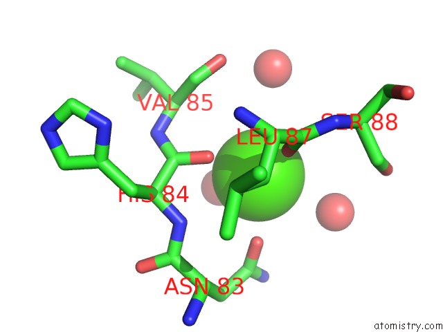
Mono view
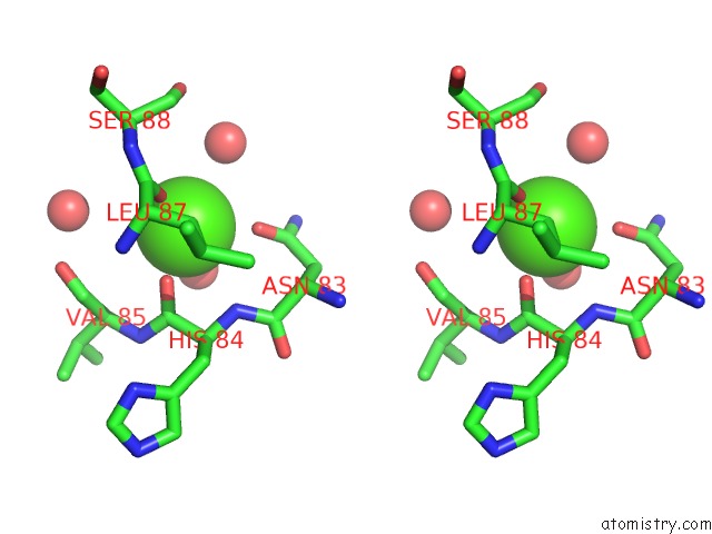
Stereo pair view
Mono view
Stereo pair view
A full contact list of Calcium with other atoms in the Ca binding
site number 1 of Crystal Structures of High Affinity Human T-Cell Receptors Bound to Pmhc Reveal Native Diagonal Binding Geometry within 5.0Å range:
|
Reference:
M.Sami,
P.J.Rizkallah,
S.Dunn,
P.Molloy,
R.Moysey,
A.Vuidepot,
E.Baston,
P.Todorov,
L.Yi,
F.Gao,
J.M.Boulter,
B.K.Jakobsen.
Crystal Structures of High Affinity Human T-Cell Receptors Bound to Peptide Major Histocompatibility Complex Reveal Native Diagonal Binding Geometry Protein Eng.Des.Sel. V. 20 397 2007.
ISSN: ISSN 1741-0126
PubMed: 17644531
DOI: 10.1093/PROTEIN/GZM033
Page generated: Tue Jul 8 07:36:21 2025
ISSN: ISSN 1741-0126
PubMed: 17644531
DOI: 10.1093/PROTEIN/GZM033
Last articles
Cl in 5K3ACl in 5K0X
Cl in 5K0W
Cl in 5K2Z
Cl in 5K2O
Cl in 5K1Z
Cl in 5K0S
Cl in 5K1X
Cl in 5K0K
Cl in 5K0T