Calcium »
PDB 2ovx-2p9k »
2p6b »
Calcium in PDB 2p6b: Crystal Structure of Human Calcineurin in Complex with Pvivit Peptide
Protein crystallography data
The structure of Crystal Structure of Human Calcineurin in Complex with Pvivit Peptide, PDB code: 2p6b
was solved by
H.Li,
L.Zhang,
A.Rao,
S.C.Harrison,
P.G.Hogan,
with X-Ray Crystallography technique. A brief refinement statistics is given in the table below:
| Resolution Low / High (Å) | 50.00 / 2.30 |
| Space group | P 21 21 21 |
| Cell size a, b, c (Å), α, β, γ (°) | 86.104, 89.155, 157.685, 90.00, 90.00, 90.00 |
| R / Rfree (%) | 19.2 / 25.4 |
Other elements in 2p6b:
The structure of Crystal Structure of Human Calcineurin in Complex with Pvivit Peptide also contains other interesting chemical elements:
| Iron | (Fe) | 2 atoms |
| Zinc | (Zn) | 2 atoms |
Calcium Binding Sites:
The binding sites of Calcium atom in the Crystal Structure of Human Calcineurin in Complex with Pvivit Peptide
(pdb code 2p6b). This binding sites where shown within
5.0 Angstroms radius around Calcium atom.
In total 8 binding sites of Calcium where determined in the Crystal Structure of Human Calcineurin in Complex with Pvivit Peptide, PDB code: 2p6b:
Jump to Calcium binding site number: 1; 2; 3; 4; 5; 6; 7; 8;
In total 8 binding sites of Calcium where determined in the Crystal Structure of Human Calcineurin in Complex with Pvivit Peptide, PDB code: 2p6b:
Jump to Calcium binding site number: 1; 2; 3; 4; 5; 6; 7; 8;
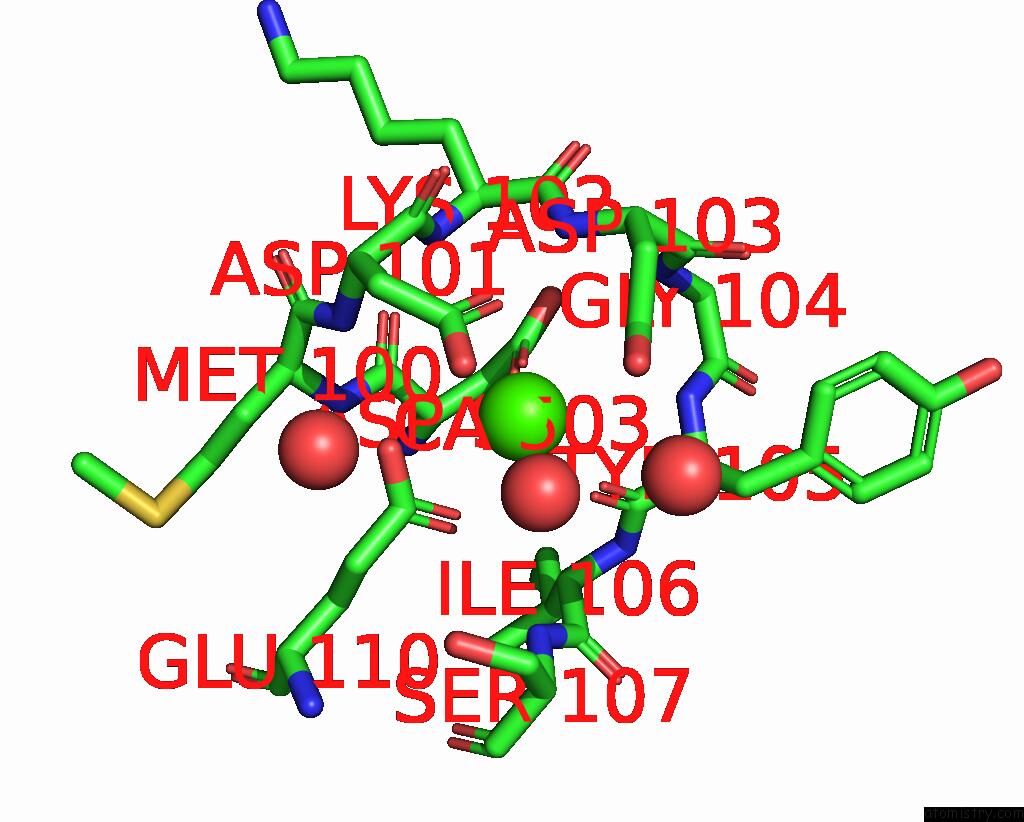
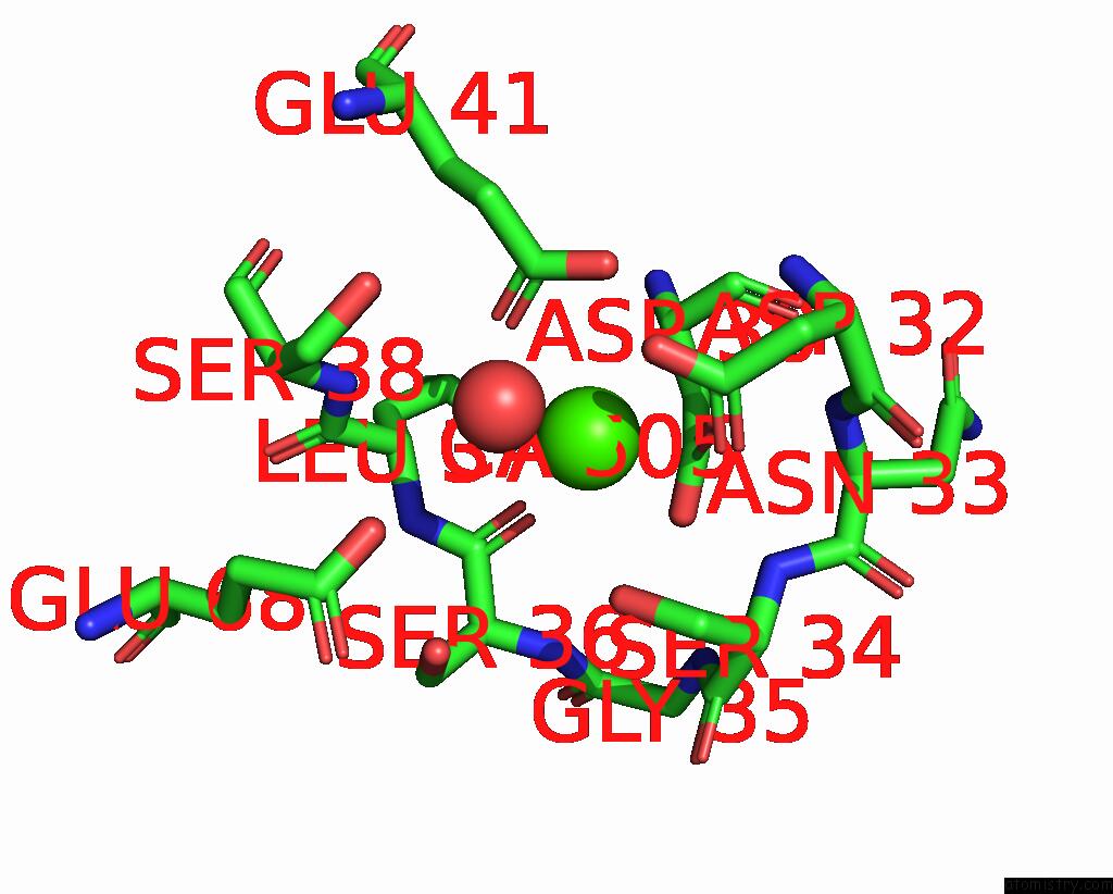
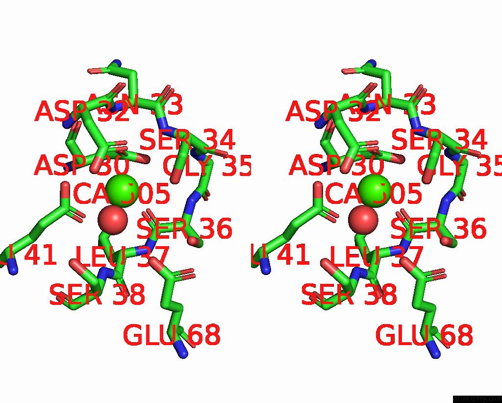
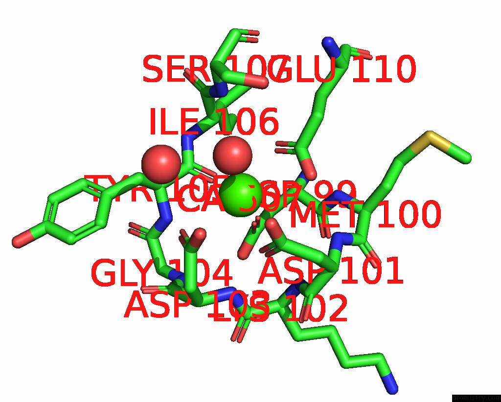
Calcium binding site 1 out of 8 in 2p6b
Go back to
Calcium binding site 1 out
of 8 in the Crystal Structure of Human Calcineurin in Complex with Pvivit Peptide
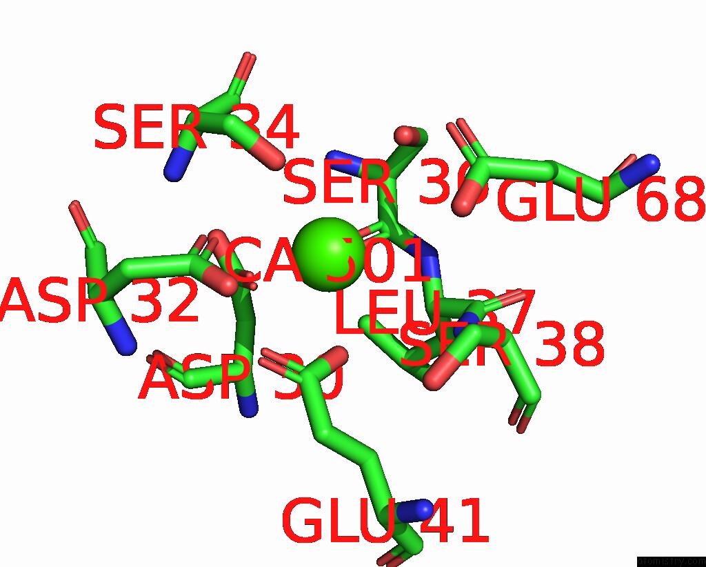
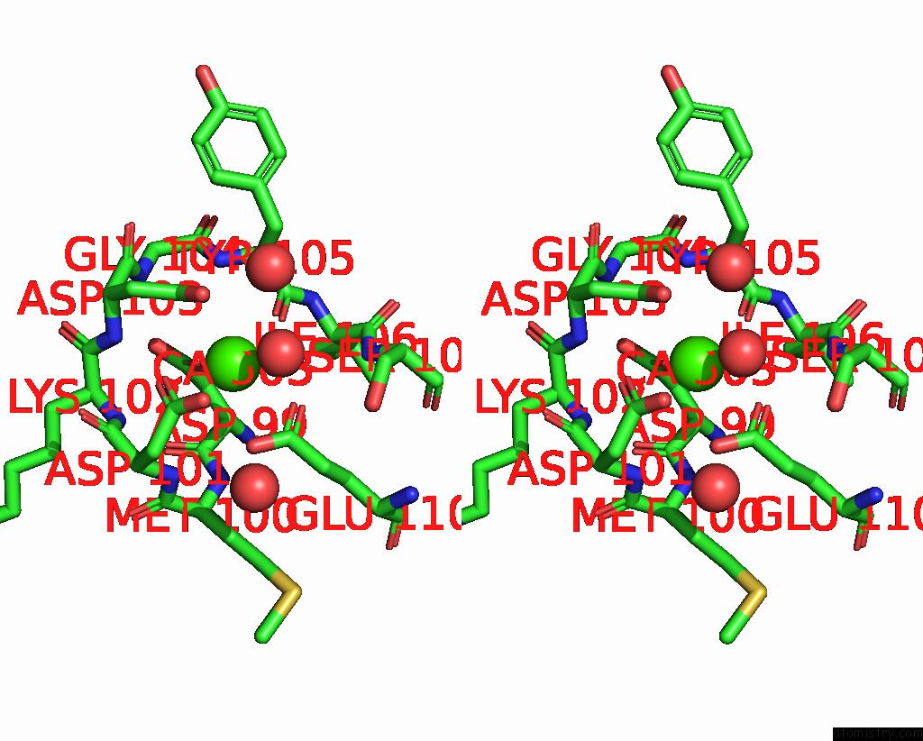
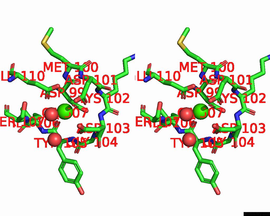
Mono view
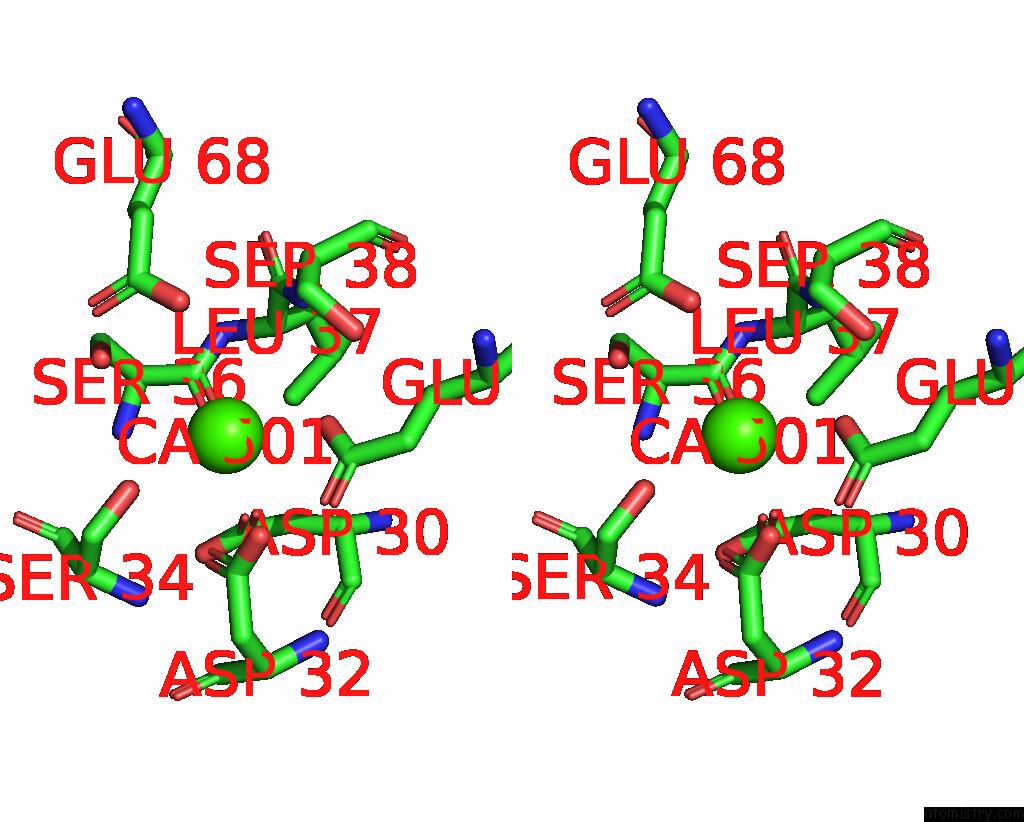
Stereo pair view
Mono view
Stereo pair view
A full contact list of Calcium with other atoms in the Ca binding
site number 1 of Crystal Structure of Human Calcineurin in Complex with Pvivit Peptide within 5.0Å range:
|
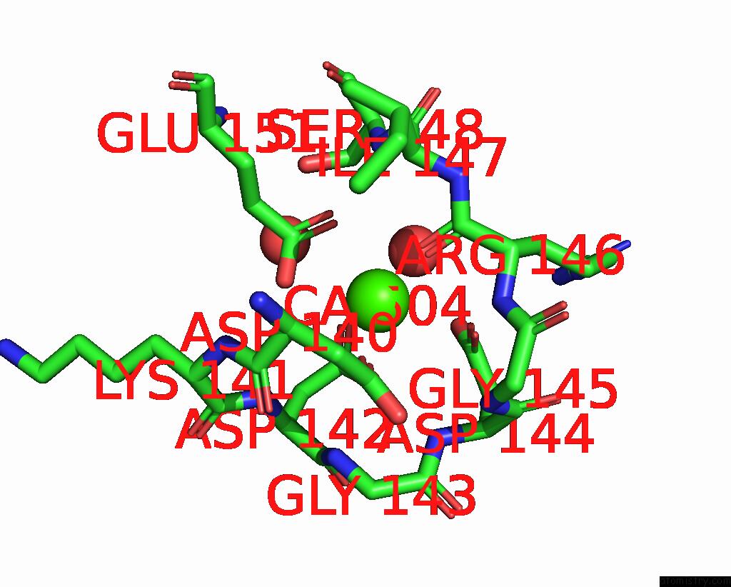
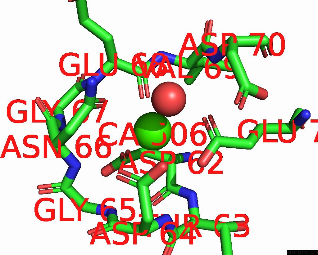
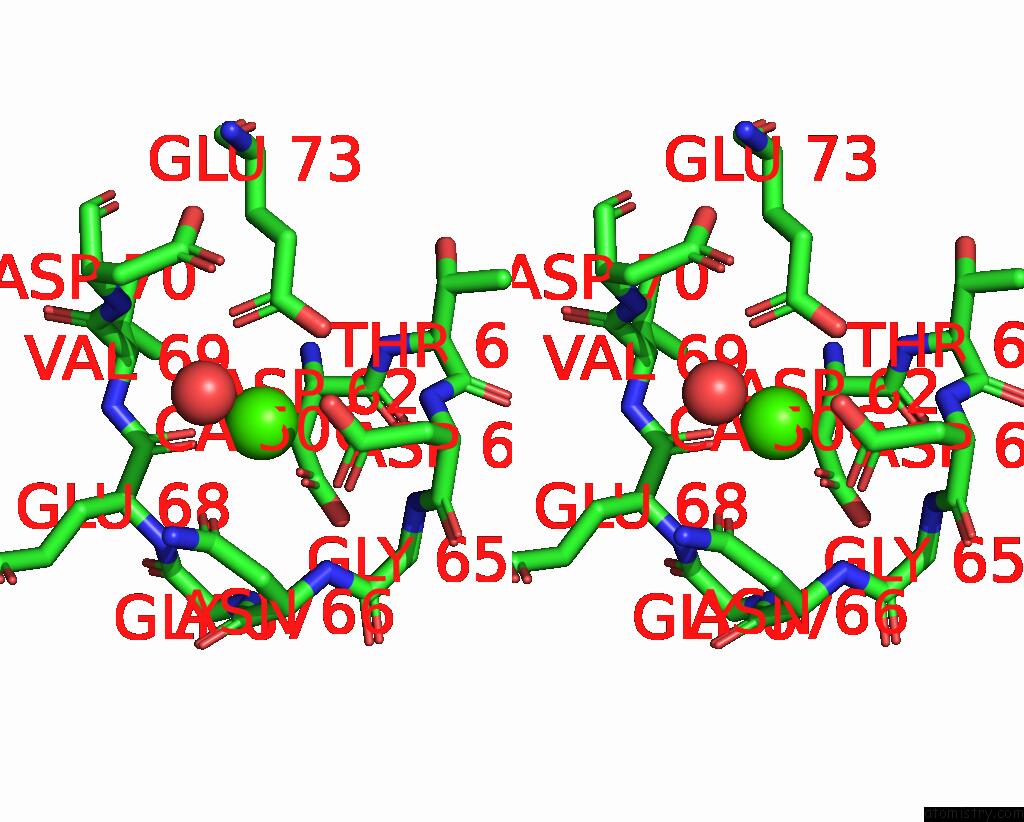
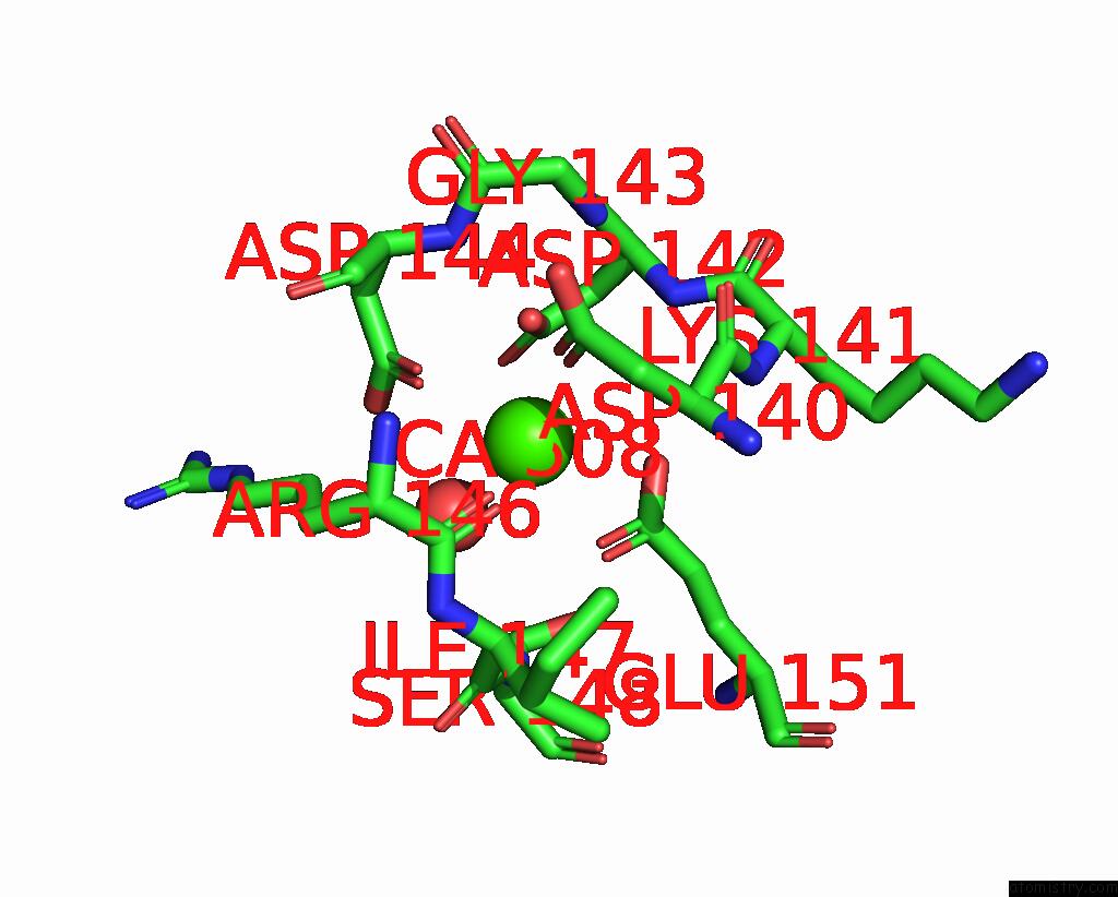
Calcium binding site 2 out of 8 in 2p6b
Go back to
Calcium binding site 2 out
of 8 in the Crystal Structure of Human Calcineurin in Complex with Pvivit Peptide
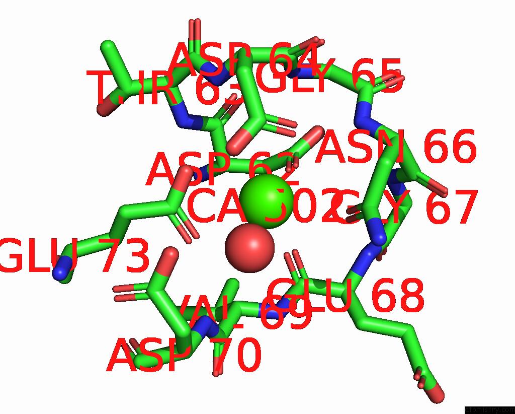
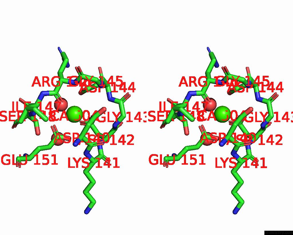
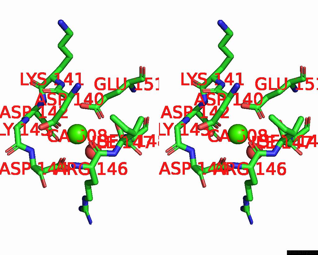
Mono view
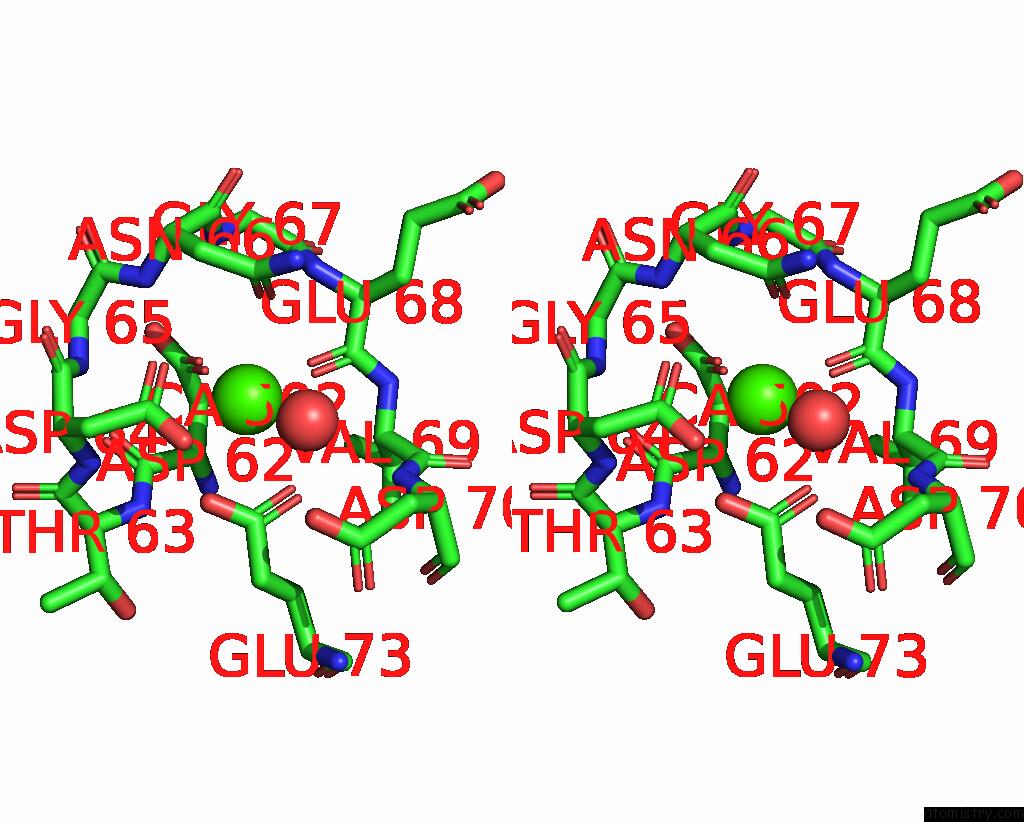
Stereo pair view
Mono view
Stereo pair view
A full contact list of Calcium with other atoms in the Ca binding
site number 2 of Crystal Structure of Human Calcineurin in Complex with Pvivit Peptide within 5.0Å range:
|
Calcium binding site 3 out of 8 in 2p6b
Go back to
Calcium binding site 3 out
of 8 in the Crystal Structure of Human Calcineurin in Complex with Pvivit Peptide
Mono view
Stereo pair view
Mono view
Stereo pair view
A full contact list of Calcium with other atoms in the Ca binding
site number 3 of Crystal Structure of Human Calcineurin in Complex with Pvivit Peptide within 5.0Å range:
|
Calcium binding site 4 out of 8 in 2p6b
Go back to
Calcium binding site 4 out
of 8 in the Crystal Structure of Human Calcineurin in Complex with Pvivit Peptide
Mono view
Stereo pair view
Mono view
Stereo pair view
A full contact list of Calcium with other atoms in the Ca binding
site number 4 of Crystal Structure of Human Calcineurin in Complex with Pvivit Peptide within 5.0Å range:
|
Calcium binding site 5 out of 8 in 2p6b
Go back to
Calcium binding site 5 out
of 8 in the Crystal Structure of Human Calcineurin in Complex with Pvivit Peptide
Mono view
Stereo pair view
Mono view
Stereo pair view
A full contact list of Calcium with other atoms in the Ca binding
site number 5 of Crystal Structure of Human Calcineurin in Complex with Pvivit Peptide within 5.0Å range:
|
Calcium binding site 6 out of 8 in 2p6b
Go back to
Calcium binding site 6 out
of 8 in the Crystal Structure of Human Calcineurin in Complex with Pvivit Peptide
Mono view
Stereo pair view
Mono view
Stereo pair view
A full contact list of Calcium with other atoms in the Ca binding
site number 6 of Crystal Structure of Human Calcineurin in Complex with Pvivit Peptide within 5.0Å range:
|
Calcium binding site 7 out of 8 in 2p6b
Go back to
Calcium binding site 7 out
of 8 in the Crystal Structure of Human Calcineurin in Complex with Pvivit Peptide
Mono view
Stereo pair view
Mono view
Stereo pair view
A full contact list of Calcium with other atoms in the Ca binding
site number 7 of Crystal Structure of Human Calcineurin in Complex with Pvivit Peptide within 5.0Å range:
|
Calcium binding site 8 out of 8 in 2p6b
Go back to
Calcium binding site 8 out
of 8 in the Crystal Structure of Human Calcineurin in Complex with Pvivit Peptide
Mono view
Stereo pair view
Mono view
Stereo pair view
A full contact list of Calcium with other atoms in the Ca binding
site number 8 of Crystal Structure of Human Calcineurin in Complex with Pvivit Peptide within 5.0Å range:
|
Reference:
H.Li,
L.Zhang,
A.Rao,
S.C.Harrison,
P.G.Hogan.
Structure of Calcineurin in Complex with Pvivit Peptide: Portrait of A Low-Affinity Signalling Interaction J.Mol.Biol. V. 369 1296 2007.
ISSN: ISSN 0022-2836
PubMed: 17498738
DOI: 10.1016/J.JMB.2007.04.032
Page generated: Fri Jul 12 15:01:08 2024
ISSN: ISSN 0022-2836
PubMed: 17498738
DOI: 10.1016/J.JMB.2007.04.032
Last articles
Zn in 9MJ5Zn in 9HNW
Zn in 9G0L
Zn in 9FNE
Zn in 9DZN
Zn in 9E0I
Zn in 9D32
Zn in 9DAK
Zn in 8ZXC
Zn in 8ZUF