Calcium »
PDB 2p9n-2prk »
2pc6 »
Calcium in PDB 2pc6: Crystal Structure of Putative Acetolactate Synthase- Small Subunit From Nitrosomonas Europaea
Enzymatic activity of Crystal Structure of Putative Acetolactate Synthase- Small Subunit From Nitrosomonas Europaea
All present enzymatic activity of Crystal Structure of Putative Acetolactate Synthase- Small Subunit From Nitrosomonas Europaea:
4.1.3.18;
4.1.3.18;
Protein crystallography data
The structure of Crystal Structure of Putative Acetolactate Synthase- Small Subunit From Nitrosomonas Europaea, PDB code: 2pc6
was solved by
J.J.Petkowski,
M.Chruszcz,
M.D.Zimmerman,
H.Zheng,
M.T.Cymborowski,
T.Skarina,
O.Onopriyenko,
A.Savchenko,
A.Edwards,
W.Minor,
A.Joachimiak,
Midwest Center For Structural Genomics (Mcsg),
with X-Ray Crystallography technique. A brief refinement statistics is given in the table below:
| Resolution Low / High (Å) | 35.58 / 2.50 |
| Space group | P 42 21 2 |
| Cell size a, b, c (Å), α, β, γ (°) | 122.123, 122.123, 111.559, 90.00, 90.00, 90.00 |
| R / Rfree (%) | 20.5 / 27.6 |
Calcium Binding Sites:
The binding sites of Calcium atom in the Crystal Structure of Putative Acetolactate Synthase- Small Subunit From Nitrosomonas Europaea
(pdb code 2pc6). This binding sites where shown within
5.0 Angstroms radius around Calcium atom.
In total 3 binding sites of Calcium where determined in the Crystal Structure of Putative Acetolactate Synthase- Small Subunit From Nitrosomonas Europaea, PDB code: 2pc6:
Jump to Calcium binding site number: 1; 2; 3;
In total 3 binding sites of Calcium where determined in the Crystal Structure of Putative Acetolactate Synthase- Small Subunit From Nitrosomonas Europaea, PDB code: 2pc6:
Jump to Calcium binding site number: 1; 2; 3;
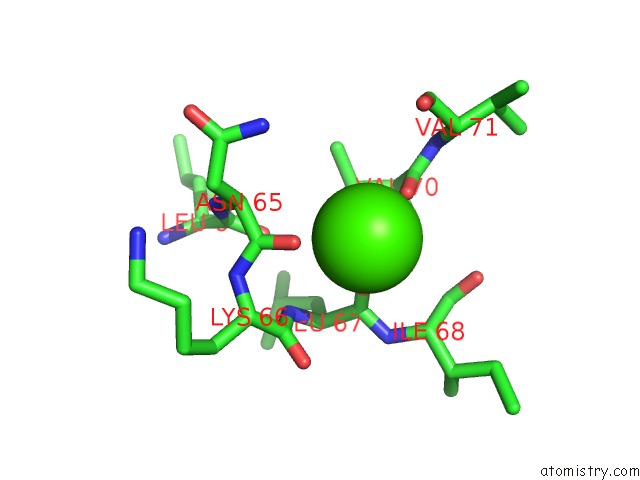
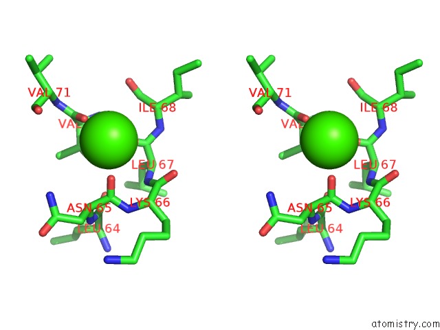
Calcium binding site 1 out of 3 in 2pc6
Go back to
Calcium binding site 1 out
of 3 in the Crystal Structure of Putative Acetolactate Synthase- Small Subunit From Nitrosomonas Europaea
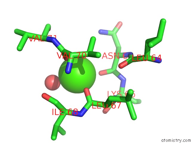
Mono view
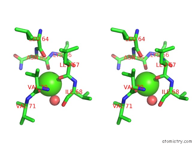
Stereo pair view
Mono view
Stereo pair view
A full contact list of Calcium with other atoms in the Ca binding
site number 1 of Crystal Structure of Putative Acetolactate Synthase- Small Subunit From Nitrosomonas Europaea within 5.0Å range:
|
Calcium binding site 2 out of 3 in 2pc6
Go back to
Calcium binding site 2 out
of 3 in the Crystal Structure of Putative Acetolactate Synthase- Small Subunit From Nitrosomonas Europaea
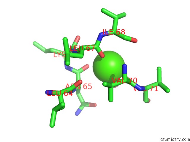
Mono view
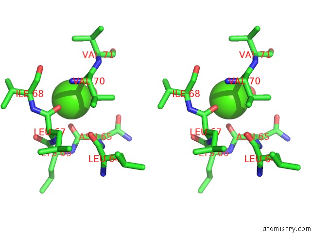
Stereo pair view
Mono view
Stereo pair view
A full contact list of Calcium with other atoms in the Ca binding
site number 2 of Crystal Structure of Putative Acetolactate Synthase- Small Subunit From Nitrosomonas Europaea within 5.0Å range:
|
Calcium binding site 3 out of 3 in 2pc6
Go back to
Calcium binding site 3 out
of 3 in the Crystal Structure of Putative Acetolactate Synthase- Small Subunit From Nitrosomonas Europaea
Mono view
Stereo pair view
Mono view
Stereo pair view
A full contact list of Calcium with other atoms in the Ca binding
site number 3 of Crystal Structure of Putative Acetolactate Synthase- Small Subunit From Nitrosomonas Europaea within 5.0Å range:
|
Reference:
J.J.Petkowski,
M.Chruszcz,
M.D.Zimmerman,
H.Zheng,
T.Skarina,
O.Onopriyenko,
M.T.Cymborowski,
K.D.Koclega,
A.Savchenko,
A.Edwards,
W.Minor.
Crystal Structures of TM0549 and NE1324--Two Orthologs of E. Coli Ahas Isozyme III Small Regulatory Subunit. Protein Sci. V. 16 1360 2007.
ISSN: ISSN 0961-8368
PubMed: 17586771
DOI: 10.1110/PS.072793807
Page generated: Fri Jul 12 15:07:38 2024
ISSN: ISSN 0961-8368
PubMed: 17586771
DOI: 10.1110/PS.072793807
Last articles
Zn in 9JYWZn in 9IR4
Zn in 9IR3
Zn in 9GMX
Zn in 9GMW
Zn in 9JEJ
Zn in 9ERF
Zn in 9ERE
Zn in 9EGV
Zn in 9EGW