Calcium »
PDB 2p9n-2prk »
2pf2 »
Calcium in PDB 2pf2: The Ca+2 Ion and Membrane Binding Structure of the Gla Domain of Ca- Prothrombin Fragment 1
Protein crystallography data
The structure of The Ca+2 Ion and Membrane Binding Structure of the Gla Domain of Ca- Prothrombin Fragment 1, PDB code: 2pf2
was solved by
M.Soriano-Garcia,
K.Padmanabhan,
A.M.De Vos,
A.Tulinsky,
with X-Ray Crystallography technique. A brief refinement statistics is given in the table below:
| Resolution Low / High (Å) | 7.00 / 2.20 |
| Space group | P 21 21 21 |
| Cell size a, b, c (Å), α, β, γ (°) | 39.390, 53.880, 129.640, 90.00, 90.00, 90.00 |
| R / Rfree (%) | n/a / n/a |
Calcium Binding Sites:
The binding sites of Calcium atom in the The Ca+2 Ion and Membrane Binding Structure of the Gla Domain of Ca- Prothrombin Fragment 1
(pdb code 2pf2). This binding sites where shown within
5.0 Angstroms radius around Calcium atom.
In total 7 binding sites of Calcium where determined in the The Ca+2 Ion and Membrane Binding Structure of the Gla Domain of Ca- Prothrombin Fragment 1, PDB code: 2pf2:
Jump to Calcium binding site number: 1; 2; 3; 4; 5; 6; 7;
In total 7 binding sites of Calcium where determined in the The Ca+2 Ion and Membrane Binding Structure of the Gla Domain of Ca- Prothrombin Fragment 1, PDB code: 2pf2:
Jump to Calcium binding site number: 1; 2; 3; 4; 5; 6; 7;
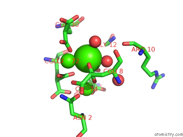
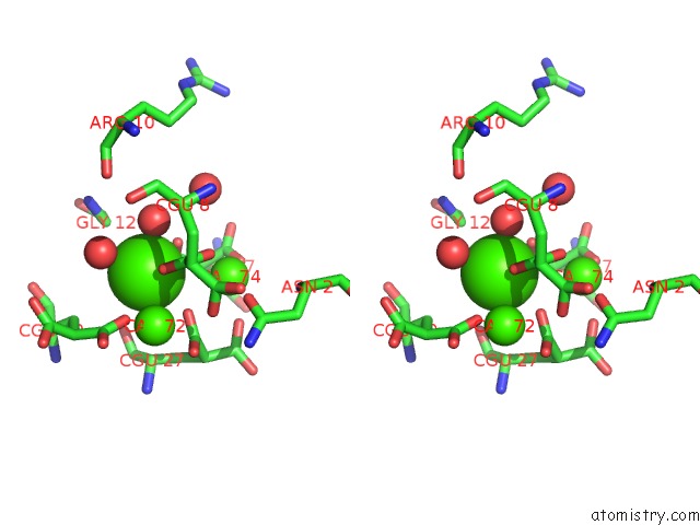
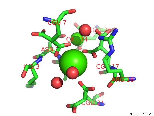
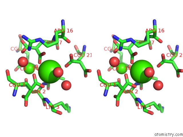
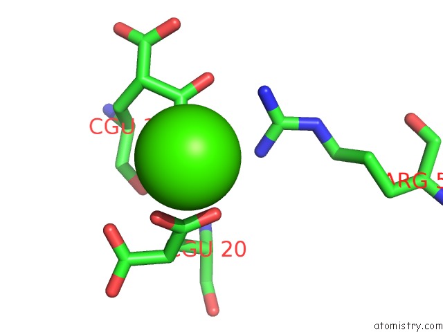
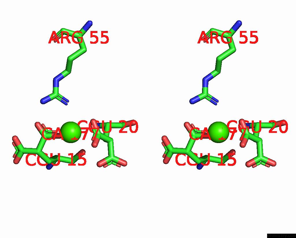
Calcium binding site 1 out of 7 in 2pf2
Go back to
Calcium binding site 1 out
of 7 in the The Ca+2 Ion and Membrane Binding Structure of the Gla Domain of Ca- Prothrombin Fragment 1
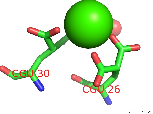
Mono view
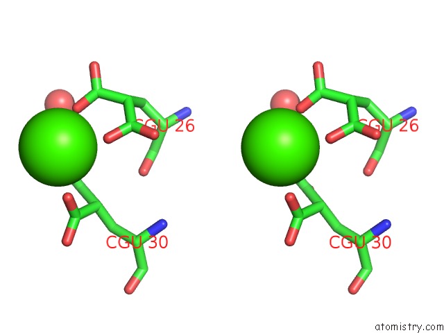
Stereo pair view
Mono view
Stereo pair view
A full contact list of Calcium with other atoms in the Ca binding
site number 1 of The Ca+2 Ion and Membrane Binding Structure of the Gla Domain of Ca- Prothrombin Fragment 1 within 5.0Å range:
|
Calcium binding site 2 out of 7 in 2pf2
Go back to
Calcium binding site 2 out
of 7 in the The Ca+2 Ion and Membrane Binding Structure of the Gla Domain of Ca- Prothrombin Fragment 1
Mono view
Stereo pair view
Mono view
Stereo pair view
A full contact list of Calcium with other atoms in the Ca binding
site number 2 of The Ca+2 Ion and Membrane Binding Structure of the Gla Domain of Ca- Prothrombin Fragment 1 within 5.0Å range:
|
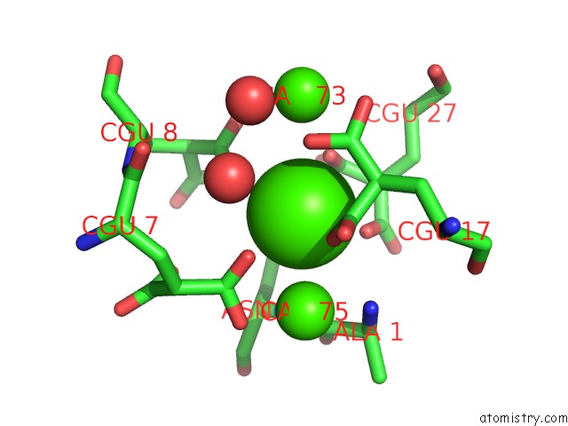
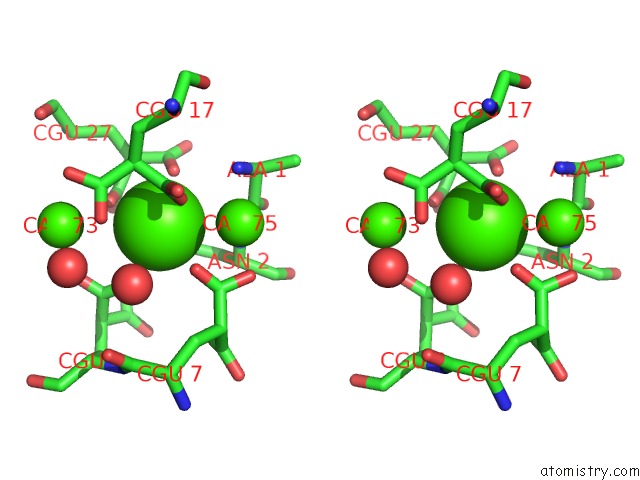
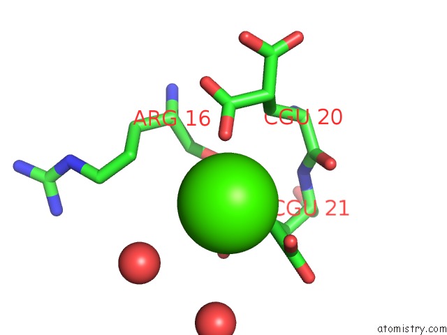
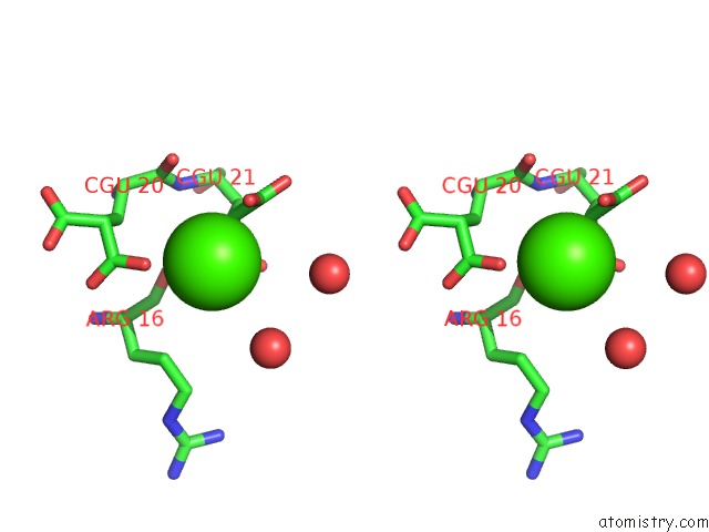
Calcium binding site 3 out of 7 in 2pf2
Go back to
Calcium binding site 3 out
of 7 in the The Ca+2 Ion and Membrane Binding Structure of the Gla Domain of Ca- Prothrombin Fragment 1
Mono view
Stereo pair view
Mono view
Stereo pair view
A full contact list of Calcium with other atoms in the Ca binding
site number 3 of The Ca+2 Ion and Membrane Binding Structure of the Gla Domain of Ca- Prothrombin Fragment 1 within 5.0Å range:
|
Calcium binding site 4 out of 7 in 2pf2
Go back to
Calcium binding site 4 out
of 7 in the The Ca+2 Ion and Membrane Binding Structure of the Gla Domain of Ca- Prothrombin Fragment 1
Mono view
Stereo pair view
Mono view
Stereo pair view
A full contact list of Calcium with other atoms in the Ca binding
site number 4 of The Ca+2 Ion and Membrane Binding Structure of the Gla Domain of Ca- Prothrombin Fragment 1 within 5.0Å range:
|
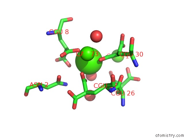
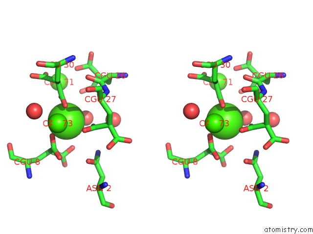
Calcium binding site 5 out of 7 in 2pf2
Go back to
Calcium binding site 5 out
of 7 in the The Ca+2 Ion and Membrane Binding Structure of the Gla Domain of Ca- Prothrombin Fragment 1
Mono view
Stereo pair view
Mono view
Stereo pair view
A full contact list of Calcium with other atoms in the Ca binding
site number 5 of The Ca+2 Ion and Membrane Binding Structure of the Gla Domain of Ca- Prothrombin Fragment 1 within 5.0Å range:
|
Calcium binding site 6 out of 7 in 2pf2
Go back to
Calcium binding site 6 out
of 7 in the The Ca+2 Ion and Membrane Binding Structure of the Gla Domain of Ca- Prothrombin Fragment 1
Mono view
Stereo pair view
Mono view
Stereo pair view
A full contact list of Calcium with other atoms in the Ca binding
site number 6 of The Ca+2 Ion and Membrane Binding Structure of the Gla Domain of Ca- Prothrombin Fragment 1 within 5.0Å range:
|
Calcium binding site 7 out of 7 in 2pf2
Go back to
Calcium binding site 7 out
of 7 in the The Ca+2 Ion and Membrane Binding Structure of the Gla Domain of Ca- Prothrombin Fragment 1
Mono view
Stereo pair view
Mono view
Stereo pair view
A full contact list of Calcium with other atoms in the Ca binding
site number 7 of The Ca+2 Ion and Membrane Binding Structure of the Gla Domain of Ca- Prothrombin Fragment 1 within 5.0Å range:
|
Reference:
M.Soriano-Garcia,
K.Padmanabhan,
A.M.De Vos,
A.Tulinsky.
The CA2+ Ion and Membrane Binding Structure of the Gla Domain of Ca-Prothrombin Fragment 1. Biochemistry V. 31 2554 1992.
ISSN: ISSN 0006-2960
PubMed: 1547238
DOI: 10.1021/BI00124A016
Page generated: Fri Jul 12 15:08:22 2024
ISSN: ISSN 0006-2960
PubMed: 1547238
DOI: 10.1021/BI00124A016
Last articles
Zn in 9MJ5Zn in 9HNW
Zn in 9G0L
Zn in 9FNE
Zn in 9DZN
Zn in 9E0I
Zn in 9D32
Zn in 9DAK
Zn in 8ZXC
Zn in 8ZUF