Calcium »
PDB 2psr-2q91 »
2pwf »
Calcium in PDB 2pwf: Crystal Structure of the Mutb D200A Mutant in Complex with Glucose
Enzymatic activity of Crystal Structure of the Mutb D200A Mutant in Complex with Glucose
All present enzymatic activity of Crystal Structure of the Mutb D200A Mutant in Complex with Glucose:
5.4.99.11;
5.4.99.11;
Protein crystallography data
The structure of Crystal Structure of the Mutb D200A Mutant in Complex with Glucose, PDB code: 2pwf
was solved by
S.Ravaud,
X.Robert,
R.Haser,
N.Aghajari,
with X-Ray Crystallography technique. A brief refinement statistics is given in the table below:
| Resolution Low / High (Å) | 45.85 / 1.80 |
| Space group | P 1 |
| Cell size a, b, c (Å), α, β, γ (°) | 63.980, 85.810, 122.140, 81.77, 81.43, 89.94 |
| R / Rfree (%) | 19.8 / 23.8 |
Calcium Binding Sites:
The binding sites of Calcium atom in the Crystal Structure of the Mutb D200A Mutant in Complex with Glucose
(pdb code 2pwf). This binding sites where shown within
5.0 Angstroms radius around Calcium atom.
In total 4 binding sites of Calcium where determined in the Crystal Structure of the Mutb D200A Mutant in Complex with Glucose, PDB code: 2pwf:
Jump to Calcium binding site number: 1; 2; 3; 4;
In total 4 binding sites of Calcium where determined in the Crystal Structure of the Mutb D200A Mutant in Complex with Glucose, PDB code: 2pwf:
Jump to Calcium binding site number: 1; 2; 3; 4;
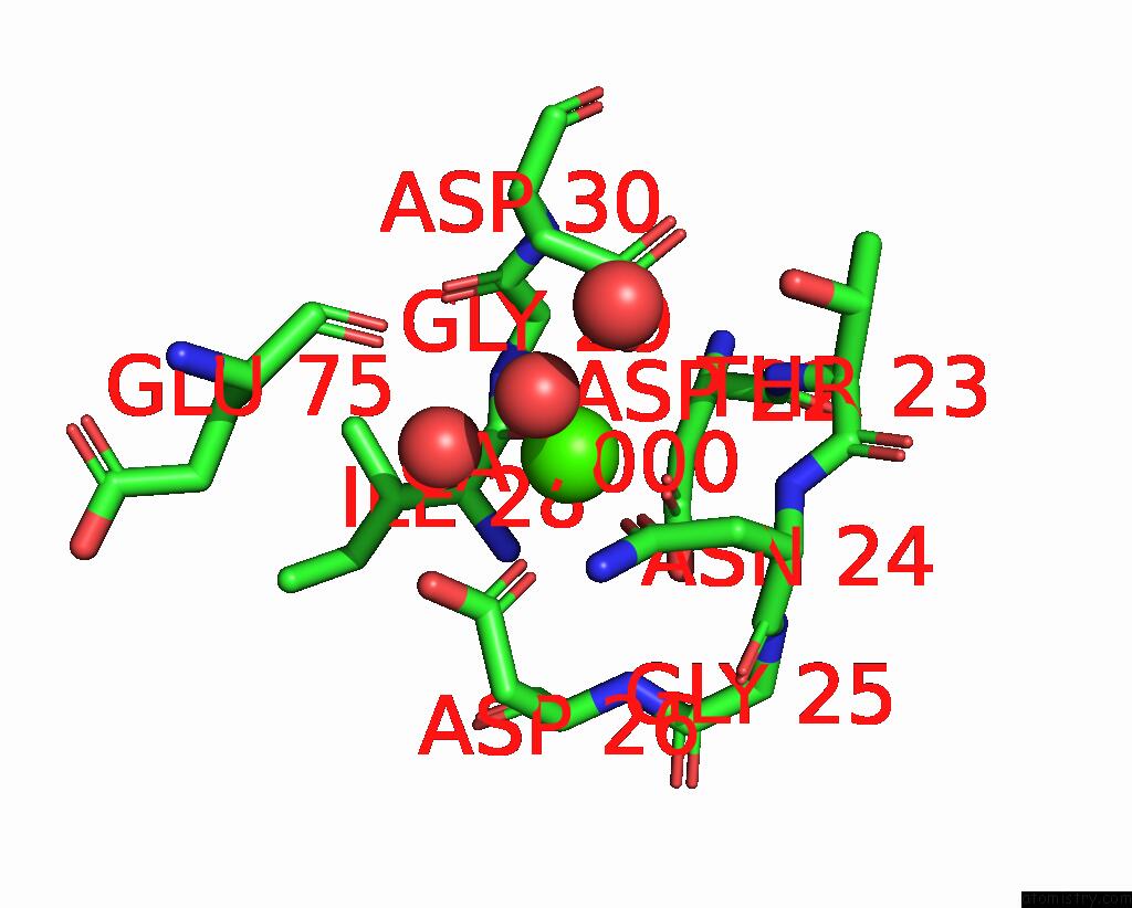
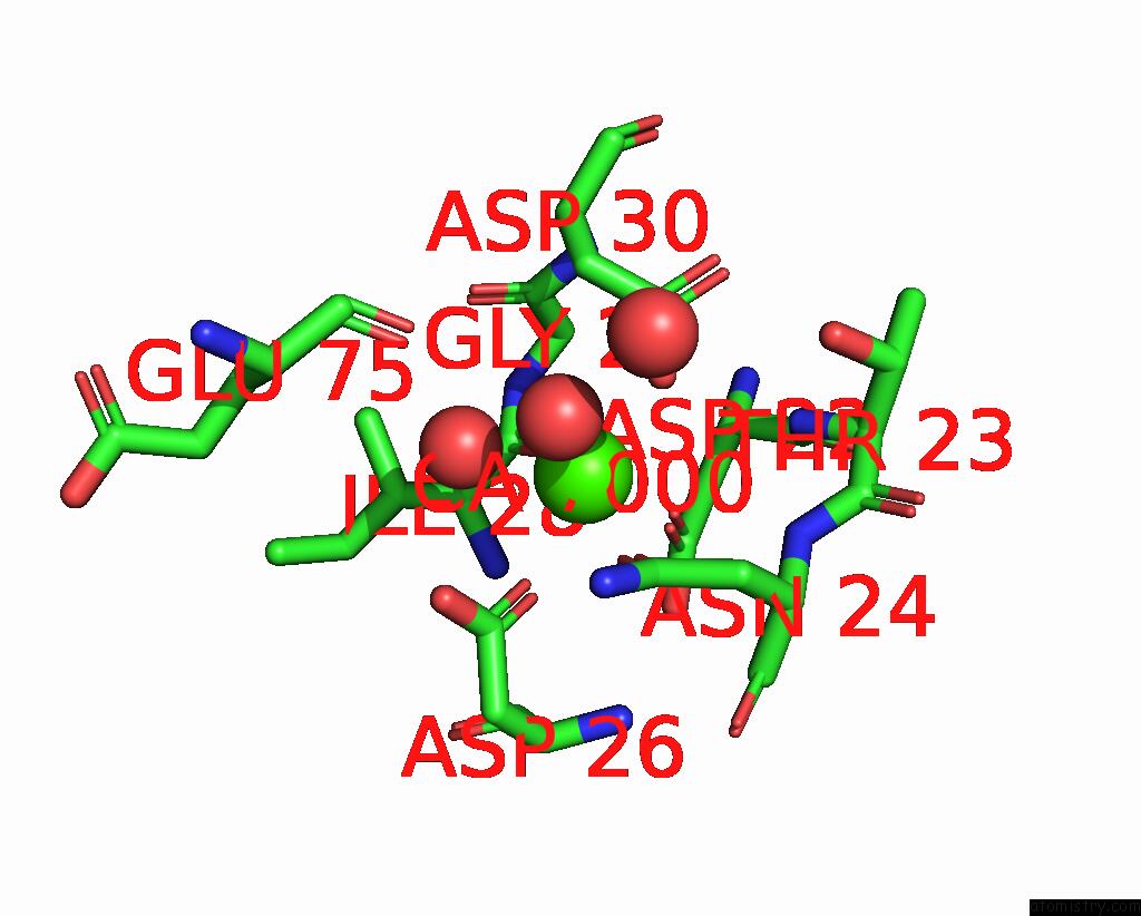
Calcium binding site 1 out of 4 in 2pwf
Go back to
Calcium binding site 1 out
of 4 in the Crystal Structure of the Mutb D200A Mutant in Complex with Glucose
Mono view
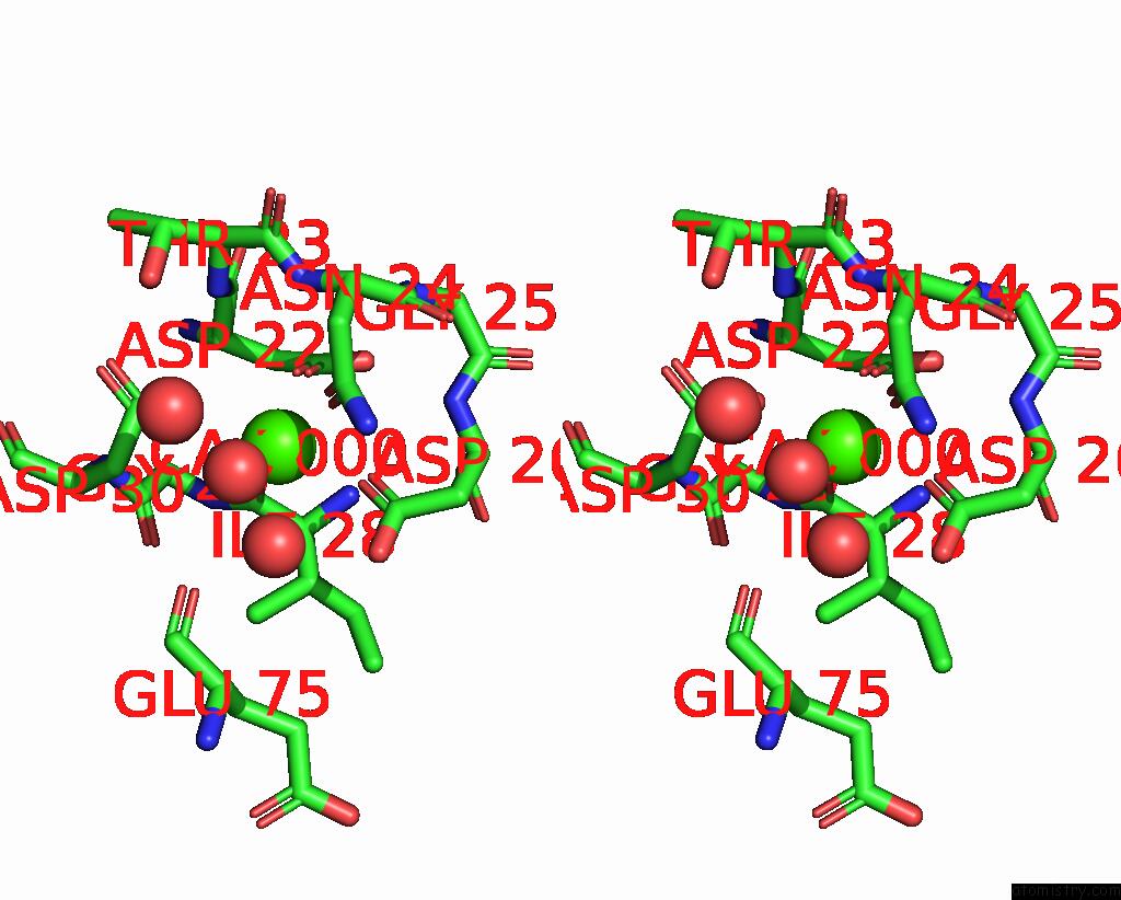
Stereo pair view
Mono view
Stereo pair view
A full contact list of Calcium with other atoms in the Ca binding
site number 1 of Crystal Structure of the Mutb D200A Mutant in Complex with Glucose within 5.0Å range:
|
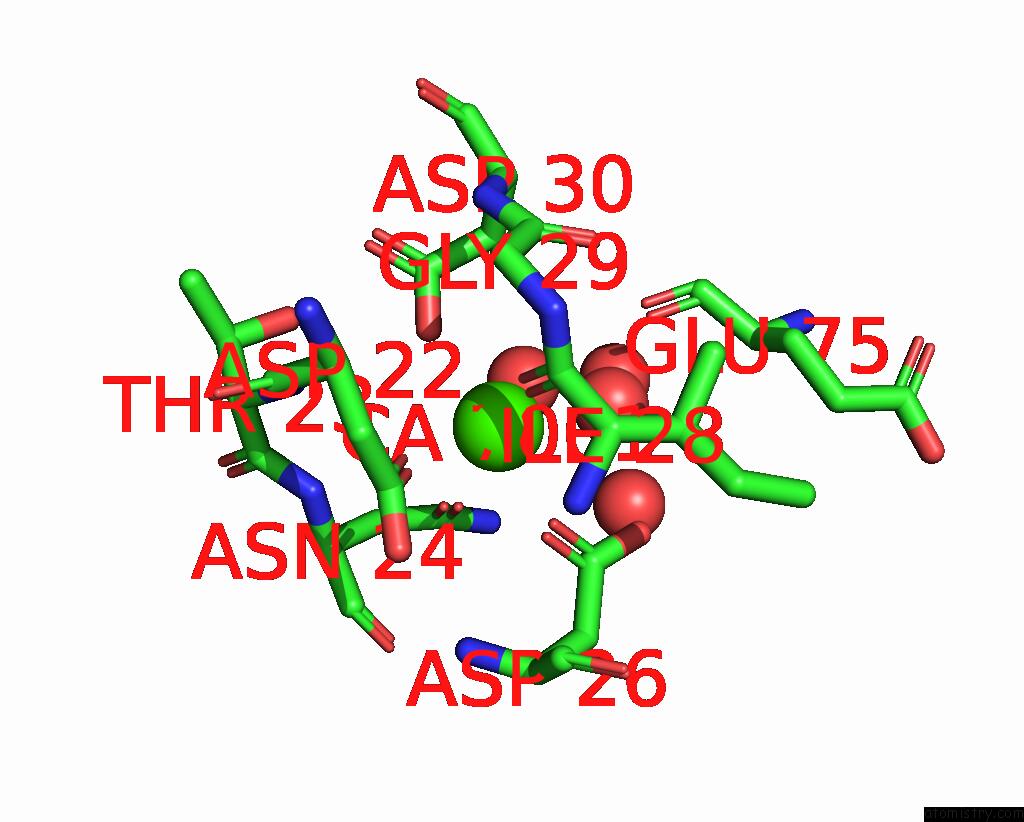
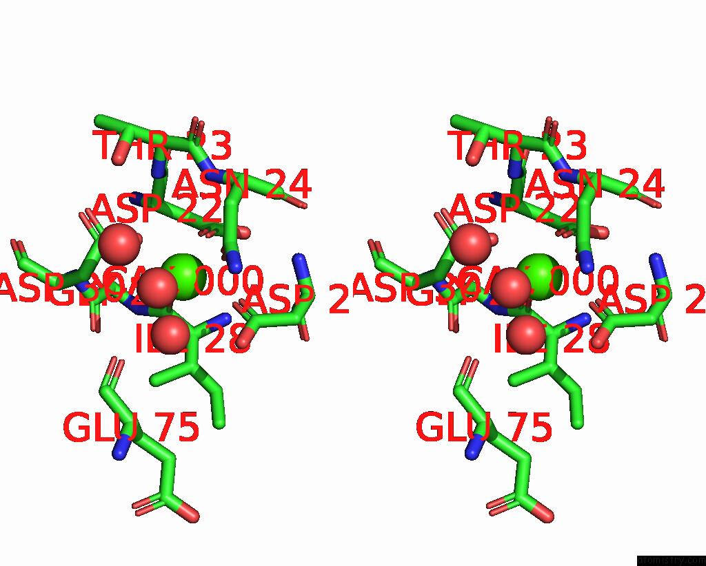
Calcium binding site 2 out of 4 in 2pwf
Go back to
Calcium binding site 2 out
of 4 in the Crystal Structure of the Mutb D200A Mutant in Complex with Glucose
Mono view
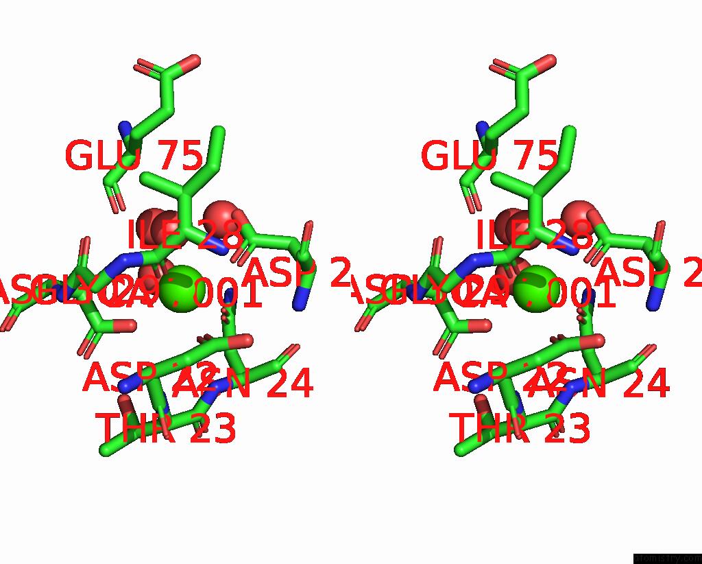
Stereo pair view
Mono view
Stereo pair view
A full contact list of Calcium with other atoms in the Ca binding
site number 2 of Crystal Structure of the Mutb D200A Mutant in Complex with Glucose within 5.0Å range:
|
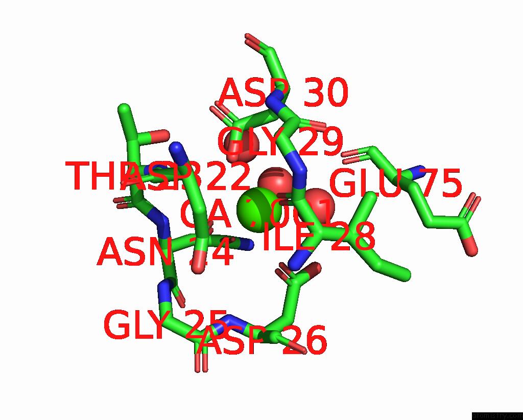
Calcium binding site 3 out of 4 in 2pwf
Go back to
Calcium binding site 3 out
of 4 in the Crystal Structure of the Mutb D200A Mutant in Complex with Glucose
Mono view
Stereo pair view
Mono view
Stereo pair view
A full contact list of Calcium with other atoms in the Ca binding
site number 3 of Crystal Structure of the Mutb D200A Mutant in Complex with Glucose within 5.0Å range:
|
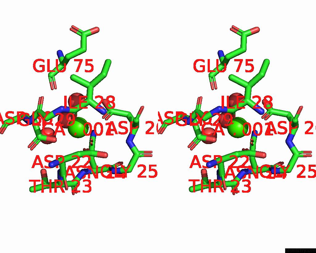
Calcium binding site 4 out of 4 in 2pwf
Go back to
Calcium binding site 4 out
of 4 in the Crystal Structure of the Mutb D200A Mutant in Complex with Glucose
Mono view
Stereo pair view
Mono view
Stereo pair view
A full contact list of Calcium with other atoms in the Ca binding
site number 4 of Crystal Structure of the Mutb D200A Mutant in Complex with Glucose within 5.0Å range:
|
Reference:
S.Ravaud,
X.Robert,
H.Watzlawick,
R.Haser,
R.Mattes,
N.Aghajari.
Trehalulose Synthase Native and Carbohydrate Complexed Structures Provide Insights Into Sucrose Isomerization. J.Biol.Chem. V. 61 100 2007.
ISSN: ISSN 0021-9258
PubMed: 17597061
DOI: 10.1074/JBC.M704515200
Page generated: Fri Jul 12 15:19:07 2024
ISSN: ISSN 0021-9258
PubMed: 17597061
DOI: 10.1074/JBC.M704515200
Last articles
Zn in 9J0NZn in 9J0O
Zn in 9J0P
Zn in 9FJX
Zn in 9EKB
Zn in 9C0F
Zn in 9CAH
Zn in 9CH0
Zn in 9CH3
Zn in 9CH1