Calcium »
PDB 2psr-2q91 »
2q1f »
Calcium in PDB 2q1f: Crystal Structure of Chondroitin Sulfate Lyase Abc From Bacteroides Thetaiotaomicron WAL2926
Enzymatic activity of Crystal Structure of Chondroitin Sulfate Lyase Abc From Bacteroides Thetaiotaomicron WAL2926
All present enzymatic activity of Crystal Structure of Chondroitin Sulfate Lyase Abc From Bacteroides Thetaiotaomicron WAL2926:
4.2.2.21;
4.2.2.21;
Protein crystallography data
The structure of Crystal Structure of Chondroitin Sulfate Lyase Abc From Bacteroides Thetaiotaomicron WAL2926, PDB code: 2q1f
was solved by
D.Shaya,
M.Cygler,
with X-Ray Crystallography technique. A brief refinement statistics is given in the table below:
| Resolution Low / High (Å) | 50.00 / 2.85 |
| Space group | P 63 |
| Cell size a, b, c (Å), α, β, γ (°) | 223.362, 223.362, 112.639, 90.00, 90.00, 120.00 |
| R / Rfree (%) | 22 / 26.2 |
Calcium Binding Sites:
The binding sites of Calcium atom in the Crystal Structure of Chondroitin Sulfate Lyase Abc From Bacteroides Thetaiotaomicron WAL2926
(pdb code 2q1f). This binding sites where shown within
5.0 Angstroms radius around Calcium atom.
In total 2 binding sites of Calcium where determined in the Crystal Structure of Chondroitin Sulfate Lyase Abc From Bacteroides Thetaiotaomicron WAL2926, PDB code: 2q1f:
Jump to Calcium binding site number: 1; 2;
In total 2 binding sites of Calcium where determined in the Crystal Structure of Chondroitin Sulfate Lyase Abc From Bacteroides Thetaiotaomicron WAL2926, PDB code: 2q1f:
Jump to Calcium binding site number: 1; 2;
Calcium binding site 1 out of 2 in 2q1f
Go back to
Calcium binding site 1 out
of 2 in the Crystal Structure of Chondroitin Sulfate Lyase Abc From Bacteroides Thetaiotaomicron WAL2926

Mono view
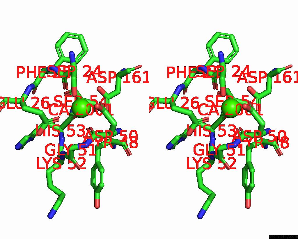
Stereo pair view
Mono view
Stereo pair view
A full contact list of Calcium with other atoms in the Ca binding
site number 1 of Crystal Structure of Chondroitin Sulfate Lyase Abc From Bacteroides Thetaiotaomicron WAL2926 within 5.0Å range:
|
Calcium binding site 2 out of 2 in 2q1f
Go back to
Calcium binding site 2 out
of 2 in the Crystal Structure of Chondroitin Sulfate Lyase Abc From Bacteroides Thetaiotaomicron WAL2926

Mono view

Stereo pair view
Mono view
Stereo pair view
A full contact list of Calcium with other atoms in the Ca binding
site number 2 of Crystal Structure of Chondroitin Sulfate Lyase Abc From Bacteroides Thetaiotaomicron WAL2926 within 5.0Å range:
|
Reference:
D.Shaya,
B.S.Hahn,
T.M.Bjerkan,
W.S.Kim,
N.Y.Park,
J.S.Sim,
Y.S.Kim,
M.Cygler.
Composite Active Site of Chondroitin Lyase Abc Accepting Both Epimers of Uronic Acid. Glycobiology V. 18 270 2008.
ISSN: ISSN 0959-6658
PubMed: 18227125
DOI: 10.1093/GLYCOB/CWN002
Page generated: Fri Jul 12 15:23:25 2024
ISSN: ISSN 0959-6658
PubMed: 18227125
DOI: 10.1093/GLYCOB/CWN002
Last articles
Zn in 9J0NZn in 9J0O
Zn in 9J0P
Zn in 9FJX
Zn in 9EKB
Zn in 9C0F
Zn in 9CAH
Zn in 9CH0
Zn in 9CH3
Zn in 9CH1