Calcium »
PDB 2psr-2q91 »
2q1n »
Calcium in PDB 2q1n: Actin Dimer Cross-Linked Between Residues 41 and 374
Protein crystallography data
The structure of Actin Dimer Cross-Linked Between Residues 41 and 374, PDB code: 2q1n
was solved by
M.R.Sawaya,
I.Pashkov,
D.S.Kudryashov,
H.Adisetiyo,
E.Reisler,
T.O.Yeates,
with X-Ray Crystallography technique. A brief refinement statistics is given in the table below:
| Resolution Low / High (Å) | 52.26 / 2.70 |
| Space group | P 1 21 1 |
| Cell size a, b, c (Å), α, β, γ (°) | 108.057, 71.813, 54.787, 90.00, 104.70, 90.00 |
| R / Rfree (%) | 22.8 / 27.7 |
Calcium Binding Sites:
The binding sites of Calcium atom in the Actin Dimer Cross-Linked Between Residues 41 and 374
(pdb code 2q1n). This binding sites where shown within
5.0 Angstroms radius around Calcium atom.
In total 4 binding sites of Calcium where determined in the Actin Dimer Cross-Linked Between Residues 41 and 374, PDB code: 2q1n:
Jump to Calcium binding site number: 1; 2; 3; 4;
In total 4 binding sites of Calcium where determined in the Actin Dimer Cross-Linked Between Residues 41 and 374, PDB code: 2q1n:
Jump to Calcium binding site number: 1; 2; 3; 4;
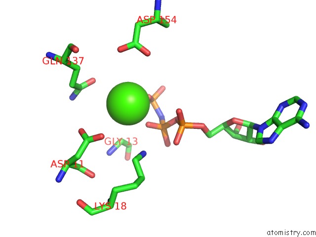
Calcium binding site 1 out of 4 in 2q1n
Go back to
Calcium binding site 1 out
of 4 in the Actin Dimer Cross-Linked Between Residues 41 and 374
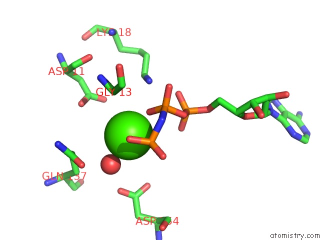
Mono view
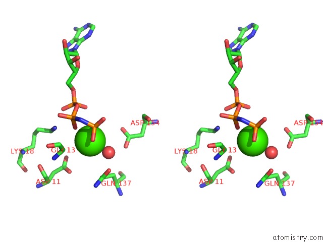
Stereo pair view
Mono view
Stereo pair view
A full contact list of Calcium with other atoms in the Ca binding
site number 1 of Actin Dimer Cross-Linked Between Residues 41 and 374 within 5.0Å range:
|
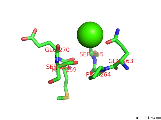
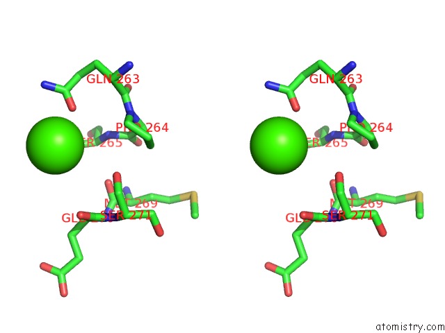
Calcium binding site 2 out of 4 in 2q1n
Go back to
Calcium binding site 2 out
of 4 in the Actin Dimer Cross-Linked Between Residues 41 and 374
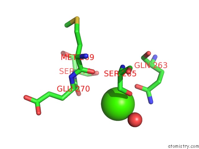
Mono view
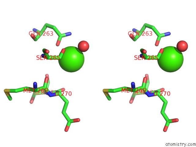
Stereo pair view
Mono view
Stereo pair view
A full contact list of Calcium with other atoms in the Ca binding
site number 2 of Actin Dimer Cross-Linked Between Residues 41 and 374 within 5.0Å range:
|
Calcium binding site 3 out of 4 in 2q1n
Go back to
Calcium binding site 3 out
of 4 in the Actin Dimer Cross-Linked Between Residues 41 and 374
Mono view

Stereo pair view
Mono view
Stereo pair view
A full contact list of Calcium with other atoms in the Ca binding
site number 3 of Actin Dimer Cross-Linked Between Residues 41 and 374 within 5.0Å range:
|
Calcium binding site 4 out of 4 in 2q1n
Go back to
Calcium binding site 4 out
of 4 in the Actin Dimer Cross-Linked Between Residues 41 and 374
Mono view
Stereo pair view
Mono view
Stereo pair view
A full contact list of Calcium with other atoms in the Ca binding
site number 4 of Actin Dimer Cross-Linked Between Residues 41 and 374 within 5.0Å range:
|
Reference:
M.R.Sawaya,
D.S.Kudryashov,
I.Pashkov,
H.Adisetiyo,
E.Reisler,
T.O.Yeates.
Multiple Crystal Structures of Actin Dimers and Their Implications For Interactions in the Actin Filament. Acta Crystallogr.,Sect.D V. 64 454 2008.
ISSN: ISSN 0907-4449
PubMed: 18391412
DOI: 10.1107/S0907444908003351
Page generated: Fri Jul 12 15:23:45 2024
ISSN: ISSN 0907-4449
PubMed: 18391412
DOI: 10.1107/S0907444908003351
Last articles
Zn in 9JYWZn in 9IR4
Zn in 9IR3
Zn in 9GMX
Zn in 9GMW
Zn in 9JEJ
Zn in 9ERF
Zn in 9ERE
Zn in 9EGV
Zn in 9EGW