Calcium »
PDB 2psr-2q91 »
2q36 »
Calcium in PDB 2q36: Actin Dimer Cross-Linked Between Residues 191 and 374 and Complexed with Kabiramide C
Protein crystallography data
The structure of Actin Dimer Cross-Linked Between Residues 191 and 374 and Complexed with Kabiramide C, PDB code: 2q36
was solved by
M.R.Sawaya,
I.Pashkov,
D.S.Kudryashov,
E.Reisler,
T.O.Yeates,
with X-Ray Crystallography technique. A brief refinement statistics is given in the table below:
| Resolution Low / High (Å) | 72.17 / 2.50 |
| Space group | P 21 21 21 |
| Cell size a, b, c (Å), α, β, γ (°) | 40.539, 74.276, 144.474, 90.00, 90.00, 90.00 |
| R / Rfree (%) | 17.5 / 22.3 |
Calcium Binding Sites:
The binding sites of Calcium atom in the Actin Dimer Cross-Linked Between Residues 191 and 374 and Complexed with Kabiramide C
(pdb code 2q36). This binding sites where shown within
5.0 Angstroms radius around Calcium atom.
In total only one binding site of Calcium was determined in the Actin Dimer Cross-Linked Between Residues 191 and 374 and Complexed with Kabiramide C, PDB code: 2q36:
In total only one binding site of Calcium was determined in the Actin Dimer Cross-Linked Between Residues 191 and 374 and Complexed with Kabiramide C, PDB code: 2q36:
Calcium binding site 1 out of 1 in 2q36
Go back to
Calcium binding site 1 out
of 1 in the Actin Dimer Cross-Linked Between Residues 191 and 374 and Complexed with Kabiramide C
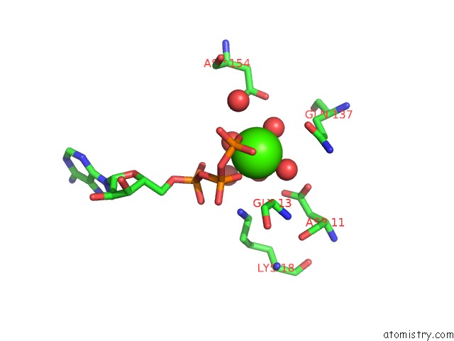
Mono view
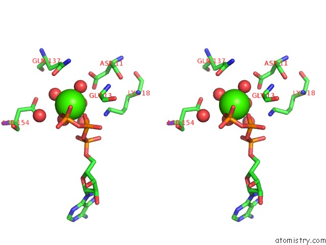
Stereo pair view
Mono view
Stereo pair view
A full contact list of Calcium with other atoms in the Ca binding
site number 1 of Actin Dimer Cross-Linked Between Residues 191 and 374 and Complexed with Kabiramide C within 5.0Å range:
|
Reference:
M.R.Sawaya,
D.S.Kudryashov,
I.Pashkov,
H.Adisetiyo,
E.Reisler,
T.O.Yeates.
Multiple Crystal Structures of Actin Dimers and Their Implications For Interactions in the Actin Filament. Acta Crystallogr.,Sect.D V. 64 454 2008.
ISSN: ISSN 0907-4449
PubMed: 18391412
DOI: 10.1107/S0907444908003351
Page generated: Tue Jul 8 07:51:52 2025
ISSN: ISSN 0907-4449
PubMed: 18391412
DOI: 10.1107/S0907444908003351
Last articles
Ca in 7LJ4Ca in 7LJ2
Ca in 7LHA
Ca in 7LG1
Ca in 7LCU
Ca in 7LFX
Ca in 7LFW
Ca in 7L8P
Ca in 7LDQ
Ca in 7LAN