Calcium »
PDB 2q97-2qwc »
2qpt »
Calcium in PDB 2qpt: Crystal Structure of An Ehd Atpase Involved in Membrane Remodelling
Protein crystallography data
The structure of Crystal Structure of An Ehd Atpase Involved in Membrane Remodelling, PDB code: 2qpt
was solved by
O.Daumke,
with X-Ray Crystallography technique. A brief refinement statistics is given in the table below:
| Resolution Low / High (Å) | 19.96 / 3.10 |
| Space group | C 1 2 1 |
| Cell size a, b, c (Å), α, β, γ (°) | 99.851, 134.653, 56.068, 90.00, 106.05, 90.00 |
| R / Rfree (%) | 23.4 / 27.6 |
Other elements in 2qpt:
The structure of Crystal Structure of An Ehd Atpase Involved in Membrane Remodelling also contains other interesting chemical elements:
| Magnesium | (Mg) | 1 atom |
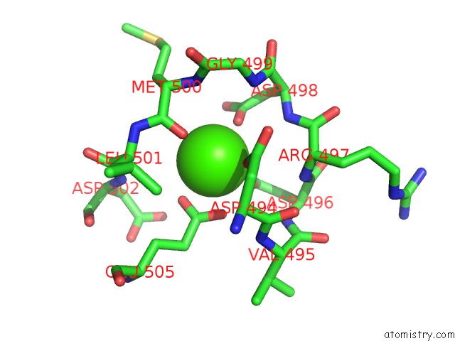
Calcium Binding Sites:
The binding sites of Calcium atom in the Crystal Structure of An Ehd Atpase Involved in Membrane Remodelling
(pdb code 2qpt). This binding sites where shown within
5.0 Angstroms radius around Calcium atom.
In total only one binding site of Calcium was determined in the Crystal Structure of An Ehd Atpase Involved in Membrane Remodelling, PDB code: 2qpt:
In total only one binding site of Calcium was determined in the Crystal Structure of An Ehd Atpase Involved in Membrane Remodelling, PDB code: 2qpt:
Calcium binding site 1 out of 1 in 2qpt
Go back to
Calcium binding site 1 out
of 1 in the Crystal Structure of An Ehd Atpase Involved in Membrane Remodelling
Mono view

Stereo pair view
Mono view
Stereo pair view
A full contact list of Calcium with other atoms in the Ca binding
site number 1 of Crystal Structure of An Ehd Atpase Involved in Membrane Remodelling within 5.0Å range:
|
Reference:
O.Daumke,
R.Lundmark,
Y.Vallis,
S.Martens,
P.J.Butler,
H.T.Mcmahon.
Architectural and Mechanistic Insights Into An Ehd Atpase Involved in Membrane Remodelling. Nature V. 449 923 2007.
ISSN: ISSN 0028-0836
PubMed: 17914359
DOI: 10.1038/NATURE06173
Page generated: Tue Jul 8 07:55:30 2025
ISSN: ISSN 0028-0836
PubMed: 17914359
DOI: 10.1038/NATURE06173
Last articles
Ca in 7G36Ca in 7G33
Ca in 7G35
Ca in 7G32
Ca in 7G34
Ca in 7G31
Ca in 7G2W
Ca in 7G30
Ca in 7G2Y
Ca in 7G2X