Calcium in PDB 2rdz: High Resolution Crystal Structure of the Escherichia Coli Cytochrome C Nitrite Reductase.
Enzymatic activity of High Resolution Crystal Structure of the Escherichia Coli Cytochrome C Nitrite Reductase.
All present enzymatic activity of High Resolution Crystal Structure of the Escherichia Coli Cytochrome C Nitrite Reductase.:
1.7.2.2;
Protein crystallography data
The structure of High Resolution Crystal Structure of the Escherichia Coli Cytochrome C Nitrite Reductase., PDB code: 2rdz
was solved by
T.A.Clarke,
A.M.Hemmings,
D.J.Richardson,
with X-Ray Crystallography technique. A brief refinement statistics is given in the table below:
Resolution Low / High (Å)
|
39.65 /
1.74
|
Space group
|
P 1 21 1
|
Cell size a, b, c (Å), α, β, γ (°)
|
90.460,
79.300,
137.580,
90.00,
101.57,
90.00
|
R / Rfree (%)
|
15.4 /
18.9
|
Other elements in 2rdz:
The structure of High Resolution Crystal Structure of the Escherichia Coli Cytochrome C Nitrite Reductase. also contains other interesting chemical elements:
Calcium Binding Sites:
The binding sites of Calcium atom in the High Resolution Crystal Structure of the Escherichia Coli Cytochrome C Nitrite Reductase.
(pdb code 2rdz). This binding sites where shown within
5.0 Angstroms radius around Calcium atom.
In total 8 binding sites of Calcium where determined in the
High Resolution Crystal Structure of the Escherichia Coli Cytochrome C Nitrite Reductase., PDB code: 2rdz:
Jump to Calcium binding site number:
1;
2;
3;
4;
5;
6;
7;
8;
Calcium binding site 1 out
of 8 in 2rdz
Go back to
Calcium Binding Sites List in 2rdz
Calcium binding site 1 out
of 8 in the High Resolution Crystal Structure of the Escherichia Coli Cytochrome C Nitrite Reductase.
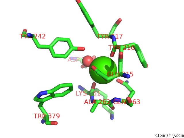
Mono view
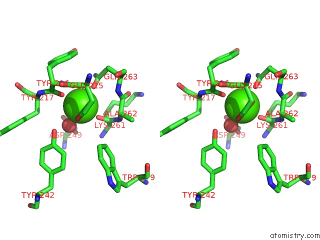
Stereo pair view
|
A full contact list of Calcium with other atoms in the Ca binding
site number 1 of High Resolution Crystal Structure of the Escherichia Coli Cytochrome C Nitrite Reductase. within 5.0Å range:
probe
|
atom
|
residue
|
distance (Å)
|
B
|
Occ
|
A:Ca1501
b:10.1
occ:1.00
|
O
|
A:LYS261
|
2.3
|
8.7
|
1.0
|
O
|
A:TYR216
|
2.4
|
10.0
|
1.0
|
O
|
A:HOH489
|
2.4
|
8.4
|
1.0
|
O
|
A:HOH499
|
2.4
|
8.7
|
1.0
|
OE1
|
A:GLN263
|
2.4
|
10.5
|
1.0
|
OE1
|
A:GLU215
|
2.5
|
9.3
|
1.0
|
OE2
|
A:GLU215
|
2.5
|
7.4
|
1.0
|
CD
|
A:GLU215
|
2.9
|
7.0
|
1.0
|
C
|
A:LYS261
|
3.5
|
9.2
|
1.0
|
C
|
A:TYR216
|
3.6
|
11.2
|
1.0
|
CD
|
A:GLN263
|
3.6
|
12.9
|
1.0
|
OH
|
A:TYR242
|
4.1
|
12.0
|
1.0
|
N
|
A:TYR216
|
4.1
|
9.3
|
1.0
|
CA
|
A:ALA262
|
4.1
|
9.0
|
1.0
|
NE2
|
A:GLN263
|
4.3
|
13.4
|
1.0
|
N
|
A:GLN263
|
4.3
|
9.2
|
1.0
|
N
|
A:ALA262
|
4.3
|
9.0
|
1.0
|
OD2
|
A:ASP249
|
4.3
|
10.9
|
1.0
|
C
|
A:ALA262
|
4.3
|
9.4
|
1.0
|
CG
|
A:GLU215
|
4.4
|
8.7
|
1.0
|
N
|
A:TYR217
|
4.5
|
10.6
|
1.0
|
CA
|
A:TYR217
|
4.5
|
12.0
|
1.0
|
CB
|
A:TYR217
|
4.5
|
12.2
|
1.0
|
CA
|
A:TYR216
|
4.5
|
10.4
|
1.0
|
CA
|
A:LYS261
|
4.6
|
8.2
|
1.0
|
OD1
|
A:ASP249
|
4.6
|
10.4
|
1.0
|
CB
|
A:LYS261
|
4.6
|
8.7
|
1.0
|
CG
|
A:ASP249
|
4.8
|
9.4
|
1.0
|
CG
|
A:GLN263
|
4.8
|
11.0
|
1.0
|
CB
|
A:GLN263
|
4.8
|
8.8
|
1.0
|
N
|
A:LYS261
|
4.9
|
8.8
|
1.0
|
CZ3
|
A:TRP379
|
4.9
|
9.6
|
1.0
|
|
Calcium binding site 2 out
of 8 in 2rdz
Go back to
Calcium Binding Sites List in 2rdz
Calcium binding site 2 out
of 8 in the High Resolution Crystal Structure of the Escherichia Coli Cytochrome C Nitrite Reductase.
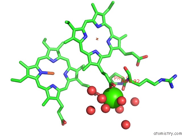
Mono view
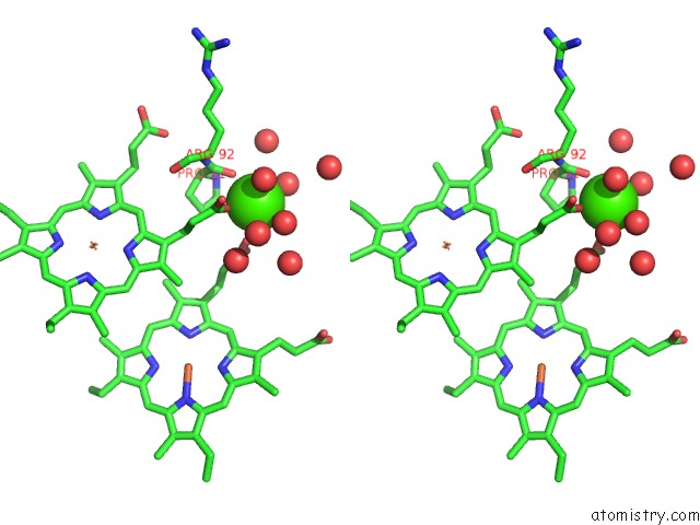
Stereo pair view
|
A full contact list of Calcium with other atoms in the Ca binding
site number 2 of High Resolution Crystal Structure of the Escherichia Coli Cytochrome C Nitrite Reductase. within 5.0Å range:
probe
|
atom
|
residue
|
distance (Å)
|
B
|
Occ
|
A:Ca1502
b:28.3
occ:1.00
|
O1A
|
A:HEC3
|
2.3
|
11.2
|
1.0
|
O1A
|
A:HEC4
|
2.4
|
26.1
|
1.0
|
O
|
A:HOH650
|
2.5
|
28.8
|
1.0
|
O
|
A:PRO91
|
2.5
|
9.8
|
1.0
|
O
|
A:HOH908
|
2.6
|
33.0
|
1.0
|
O
|
A:HOH936
|
2.6
|
33.4
|
1.0
|
O
|
A:HOH623
|
2.8
|
33.4
|
1.0
|
CGA
|
A:HEC3
|
3.4
|
13.6
|
1.0
|
CGA
|
A:HEC4
|
3.5
|
24.6
|
1.0
|
C
|
A:PRO91
|
3.6
|
9.7
|
1.0
|
O2A
|
A:HEC3
|
4.0
|
13.4
|
1.0
|
O2A
|
A:HEC4
|
4.0
|
26.4
|
1.0
|
CA
|
A:ARG92
|
4.2
|
9.5
|
1.0
|
O
|
A:HOH556
|
4.3
|
19.1
|
1.0
|
N
|
A:ARG92
|
4.3
|
9.4
|
1.0
|
O
|
A:HOH898
|
4.3
|
37.0
|
1.0
|
O
|
A:HOH721
|
4.5
|
26.9
|
1.0
|
CA
|
A:PRO91
|
4.6
|
9.4
|
1.0
|
CAA
|
A:HEC3
|
4.6
|
8.7
|
1.0
|
CG
|
A:PRO91
|
4.6
|
10.1
|
1.0
|
CBA
|
A:HEC3
|
4.6
|
8.5
|
1.0
|
N
|
A:PRO91
|
4.6
|
9.6
|
1.0
|
CBA
|
A:HEC4
|
4.7
|
18.5
|
1.0
|
O
|
A:HOH747
|
4.7
|
28.1
|
1.0
|
CB
|
A:PRO91
|
4.8
|
10.3
|
1.0
|
CD
|
A:PRO91
|
4.8
|
9.4
|
1.0
|
O
|
A:HOH814
|
4.9
|
39.6
|
1.0
|
CB
|
A:ARG92
|
5.0
|
8.5
|
1.0
|
|
Calcium binding site 3 out
of 8 in 2rdz
Go back to
Calcium Binding Sites List in 2rdz
Calcium binding site 3 out
of 8 in the High Resolution Crystal Structure of the Escherichia Coli Cytochrome C Nitrite Reductase.
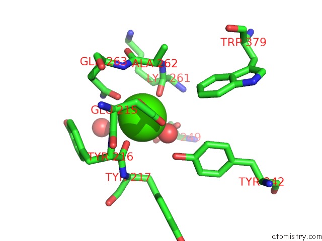
Mono view
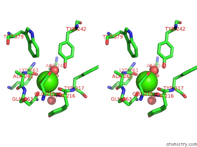
Stereo pair view
|
A full contact list of Calcium with other atoms in the Ca binding
site number 3 of High Resolution Crystal Structure of the Escherichia Coli Cytochrome C Nitrite Reductase. within 5.0Å range:
probe
|
atom
|
residue
|
distance (Å)
|
B
|
Occ
|
B:Ca1501
b:10.6
occ:1.00
|
O
|
B:TYR216
|
2.4
|
8.7
|
1.0
|
O
|
B:LYS261
|
2.4
|
9.0
|
1.0
|
O
|
B:HOH504
|
2.4
|
10.4
|
1.0
|
O
|
B:HOH528
|
2.4
|
9.2
|
1.0
|
OE1
|
B:GLN263
|
2.4
|
11.8
|
1.0
|
OE1
|
B:GLU215
|
2.5
|
8.7
|
1.0
|
OE2
|
B:GLU215
|
2.6
|
9.6
|
1.0
|
CD
|
B:GLU215
|
2.9
|
8.4
|
1.0
|
C
|
B:LYS261
|
3.5
|
9.2
|
1.0
|
C
|
B:TYR216
|
3.6
|
9.1
|
1.0
|
CD
|
B:GLN263
|
3.6
|
12.6
|
1.0
|
OH
|
B:TYR242
|
4.0
|
12.2
|
1.0
|
N
|
B:TYR216
|
4.1
|
8.1
|
1.0
|
NE2
|
B:GLN263
|
4.1
|
11.0
|
1.0
|
CA
|
B:ALA262
|
4.1
|
9.3
|
1.0
|
N
|
B:GLN263
|
4.2
|
9.2
|
1.0
|
N
|
B:ALA262
|
4.3
|
10.1
|
1.0
|
C
|
B:ALA262
|
4.3
|
9.9
|
1.0
|
OD2
|
B:ASP249
|
4.4
|
11.0
|
1.0
|
CG
|
B:GLU215
|
4.4
|
7.2
|
1.0
|
N
|
B:TYR217
|
4.5
|
9.7
|
1.0
|
CA
|
B:TYR216
|
4.5
|
8.6
|
1.0
|
CA
|
B:TYR217
|
4.5
|
10.7
|
1.0
|
OD1
|
B:ASP249
|
4.6
|
11.3
|
1.0
|
CA
|
B:LYS261
|
4.6
|
9.4
|
1.0
|
CB
|
B:TYR217
|
4.6
|
11.3
|
1.0
|
CB
|
B:LYS261
|
4.7
|
8.9
|
1.0
|
O
|
B:HOH656
|
4.8
|
28.7
|
1.0
|
CG
|
B:GLN263
|
4.8
|
9.7
|
1.0
|
CG
|
B:ASP249
|
4.8
|
11.4
|
1.0
|
CB
|
B:GLN263
|
4.9
|
9.4
|
1.0
|
N
|
B:LYS261
|
4.9
|
9.6
|
1.0
|
CZ3
|
B:TRP379
|
4.9
|
8.8
|
1.0
|
CZ
|
B:TYR242
|
5.0
|
12.5
|
1.0
|
|
Calcium binding site 4 out
of 8 in 2rdz
Go back to
Calcium Binding Sites List in 2rdz
Calcium binding site 4 out
of 8 in the High Resolution Crystal Structure of the Escherichia Coli Cytochrome C Nitrite Reductase.
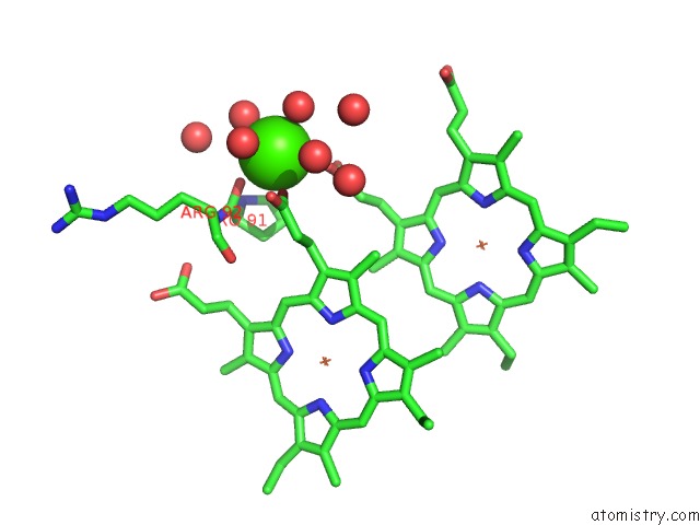
Mono view
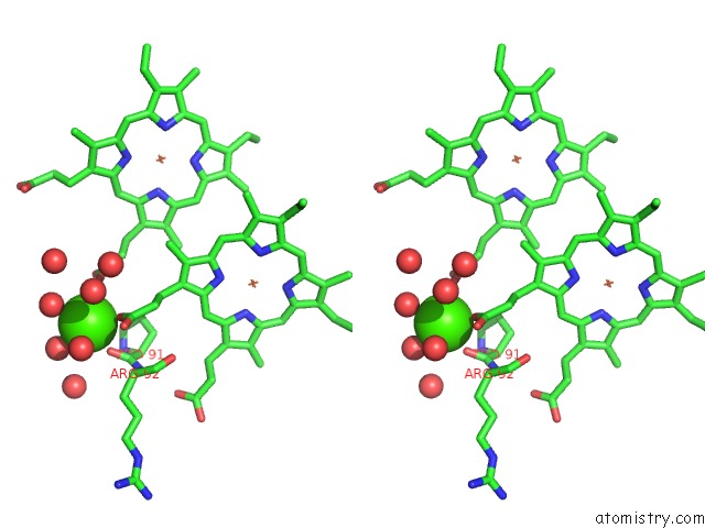
Stereo pair view
|
A full contact list of Calcium with other atoms in the Ca binding
site number 4 of High Resolution Crystal Structure of the Escherichia Coli Cytochrome C Nitrite Reductase. within 5.0Å range:
probe
|
atom
|
residue
|
distance (Å)
|
B
|
Occ
|
B:Ca1502
b:25.8
occ:1.00
|
O1A
|
B:HEC3
|
2.4
|
11.5
|
1.0
|
O
|
B:HOH676
|
2.4
|
27.0
|
1.0
|
O
|
B:PRO91
|
2.4
|
8.7
|
1.0
|
O
|
B:HOH716
|
2.5
|
31.3
|
1.0
|
O
|
B:HOH955
|
2.6
|
32.7
|
1.0
|
O1A
|
B:HEC4
|
2.6
|
25.5
|
1.0
|
O
|
B:HOH661
|
3.0
|
31.2
|
1.0
|
CGA
|
B:HEC3
|
3.5
|
11.7
|
1.0
|
C
|
B:PRO91
|
3.5
|
9.0
|
1.0
|
CGA
|
B:HEC4
|
3.6
|
23.0
|
1.0
|
O2A
|
B:HEC4
|
3.9
|
23.3
|
1.0
|
O2A
|
B:HEC3
|
4.0
|
11.9
|
1.0
|
CA
|
B:ARG92
|
4.2
|
8.3
|
1.0
|
O
|
B:HOH525
|
4.3
|
13.0
|
1.0
|
N
|
B:ARG92
|
4.3
|
8.2
|
1.0
|
O
|
B:HOH902
|
4.5
|
29.1
|
1.0
|
CA
|
B:PRO91
|
4.5
|
9.4
|
1.0
|
N
|
B:PRO91
|
4.5
|
9.6
|
1.0
|
O
|
B:HOH658
|
4.5
|
22.5
|
1.0
|
CD
|
B:PRO91
|
4.7
|
10.3
|
1.0
|
CG
|
B:PRO91
|
4.7
|
11.0
|
1.0
|
CAA
|
B:HEC3
|
4.7
|
7.8
|
1.0
|
CBA
|
B:HEC3
|
4.7
|
10.2
|
1.0
|
CBA
|
B:HEC4
|
4.8
|
17.8
|
1.0
|
CB
|
B:PRO91
|
4.8
|
9.7
|
1.0
|
O
|
B:HOH720
|
4.8
|
34.6
|
1.0
|
|
Calcium binding site 5 out
of 8 in 2rdz
Go back to
Calcium Binding Sites List in 2rdz
Calcium binding site 5 out
of 8 in the High Resolution Crystal Structure of the Escherichia Coli Cytochrome C Nitrite Reductase.
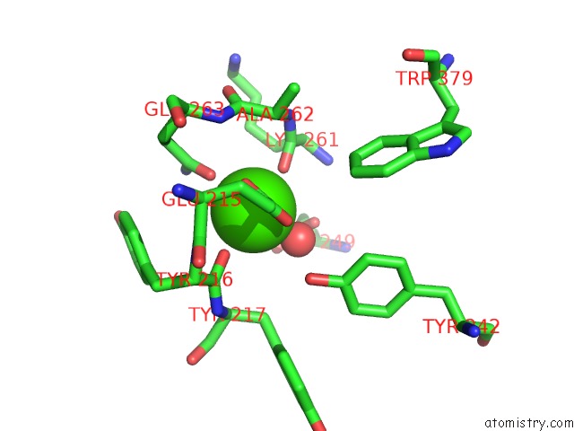
Mono view
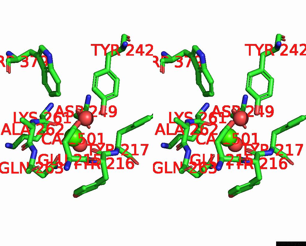
Stereo pair view
|
A full contact list of Calcium with other atoms in the Ca binding
site number 5 of High Resolution Crystal Structure of the Escherichia Coli Cytochrome C Nitrite Reductase. within 5.0Å range:
probe
|
atom
|
residue
|
distance (Å)
|
B
|
Occ
|
C:Ca1501
b:11.3
occ:1.00
|
O
|
C:TYR216
|
2.3
|
11.6
|
1.0
|
O
|
C:LYS261
|
2.4
|
10.8
|
1.0
|
O
|
C:HOH495
|
2.4
|
9.8
|
1.0
|
OE1
|
C:GLN263
|
2.4
|
13.0
|
1.0
|
O
|
C:HOH514
|
2.4
|
12.1
|
1.0
|
OE1
|
C:GLU215
|
2.5
|
10.2
|
1.0
|
OE2
|
C:GLU215
|
2.6
|
12.2
|
1.0
|
CD
|
C:GLU215
|
2.9
|
11.3
|
1.0
|
CD
|
C:GLN263
|
3.5
|
14.3
|
1.0
|
C
|
C:LYS261
|
3.6
|
10.6
|
1.0
|
C
|
C:TYR216
|
3.6
|
13.1
|
1.0
|
OH
|
C:TYR242
|
4.0
|
13.6
|
1.0
|
NE2
|
C:GLN263
|
4.1
|
15.8
|
1.0
|
N
|
C:TYR216
|
4.1
|
11.7
|
1.0
|
CA
|
C:ALA262
|
4.2
|
9.9
|
1.0
|
OD2
|
C:ASP249
|
4.3
|
12.3
|
1.0
|
N
|
C:ALA262
|
4.3
|
10.2
|
1.0
|
N
|
C:GLN263
|
4.4
|
10.6
|
1.0
|
CG
|
C:GLU215
|
4.4
|
10.9
|
1.0
|
C
|
C:ALA262
|
4.4
|
10.8
|
1.0
|
N
|
C:TYR217
|
4.5
|
12.9
|
1.0
|
CA
|
C:TYR216
|
4.5
|
12.3
|
1.0
|
CA
|
C:TYR217
|
4.5
|
13.8
|
1.0
|
OD1
|
C:ASP249
|
4.6
|
11.9
|
1.0
|
CA
|
C:LYS261
|
4.6
|
10.3
|
1.0
|
CB
|
C:TYR217
|
4.6
|
14.0
|
1.0
|
CB
|
C:LYS261
|
4.7
|
10.6
|
1.0
|
CG
|
C:ASP249
|
4.7
|
12.3
|
1.0
|
CG
|
C:GLN263
|
4.8
|
10.4
|
1.0
|
N
|
C:LYS261
|
4.9
|
10.5
|
1.0
|
CB
|
C:GLN263
|
4.9
|
10.4
|
1.0
|
CZ3
|
C:TRP379
|
4.9
|
10.3
|
1.0
|
CZ
|
C:TYR242
|
5.0
|
12.5
|
1.0
|
|
Calcium binding site 6 out
of 8 in 2rdz
Go back to
Calcium Binding Sites List in 2rdz
Calcium binding site 6 out
of 8 in the High Resolution Crystal Structure of the Escherichia Coli Cytochrome C Nitrite Reductase.
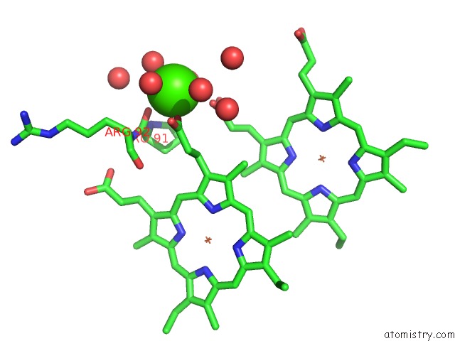
Mono view
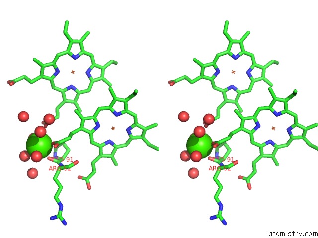
Stereo pair view
|
A full contact list of Calcium with other atoms in the Ca binding
site number 6 of High Resolution Crystal Structure of the Escherichia Coli Cytochrome C Nitrite Reductase. within 5.0Å range:
probe
|
atom
|
residue
|
distance (Å)
|
B
|
Occ
|
C:Ca1502
b:31.9
occ:1.00
|
O1A
|
C:HEC3
|
2.3
|
14.1
|
1.0
|
O
|
C:PRO91
|
2.4
|
13.2
|
1.0
|
O
|
C:HOH609
|
2.6
|
30.6
|
1.0
|
O
|
C:HOH769
|
2.7
|
38.4
|
1.0
|
O1A
|
C:HEC4
|
2.8
|
27.5
|
1.0
|
O
|
C:HOH712
|
2.8
|
32.6
|
1.0
|
CGA
|
C:HEC3
|
3.5
|
15.8
|
1.0
|
C
|
C:PRO91
|
3.5
|
13.8
|
1.0
|
CGA
|
C:HEC4
|
3.6
|
25.3
|
1.0
|
O2A
|
C:HEC4
|
4.0
|
28.0
|
1.0
|
O2A
|
C:HEC3
|
4.0
|
15.5
|
1.0
|
CA
|
C:ARG92
|
4.2
|
13.5
|
1.0
|
O
|
C:HOH568
|
4.2
|
19.1
|
1.0
|
N
|
C:ARG92
|
4.3
|
13.4
|
1.0
|
O
|
C:HOH660
|
4.4
|
29.4
|
1.0
|
O
|
C:HOH718
|
4.5
|
39.5
|
1.0
|
CA
|
C:PRO91
|
4.5
|
13.3
|
1.0
|
N
|
C:PRO91
|
4.6
|
13.5
|
1.0
|
CG
|
C:PRO91
|
4.6
|
13.7
|
1.0
|
CAA
|
C:HEC3
|
4.6
|
12.9
|
1.0
|
CBA
|
C:HEC3
|
4.6
|
14.6
|
1.0
|
CD
|
C:PRO91
|
4.8
|
13.6
|
1.0
|
O
|
C:HOH640
|
4.8
|
30.0
|
1.0
|
CB
|
C:PRO91
|
4.8
|
13.9
|
1.0
|
CBA
|
C:HEC4
|
4.8
|
20.6
|
1.0
|
CB
|
C:ARG92
|
5.0
|
12.8
|
1.0
|
|
Calcium binding site 7 out
of 8 in 2rdz
Go back to
Calcium Binding Sites List in 2rdz
Calcium binding site 7 out
of 8 in the High Resolution Crystal Structure of the Escherichia Coli Cytochrome C Nitrite Reductase.
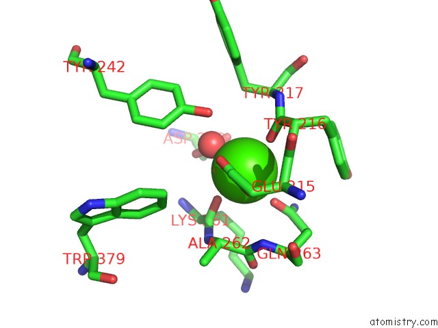
Mono view
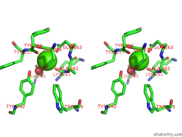
Stereo pair view
|
A full contact list of Calcium with other atoms in the Ca binding
site number 7 of High Resolution Crystal Structure of the Escherichia Coli Cytochrome C Nitrite Reductase. within 5.0Å range:
probe
|
atom
|
residue
|
distance (Å)
|
B
|
Occ
|
D:Ca1501
b:8.8
occ:1.00
|
O
|
D:TYR216
|
2.3
|
6.8
|
1.0
|
O
|
D:LYS261
|
2.3
|
7.8
|
1.0
|
O
|
D:HOH497
|
2.4
|
6.2
|
1.0
|
OE1
|
D:GLN263
|
2.4
|
11.6
|
1.0
|
O
|
D:HOH520
|
2.4
|
7.1
|
1.0
|
OE2
|
D:GLU215
|
2.5
|
7.2
|
1.0
|
OE1
|
D:GLU215
|
2.5
|
7.5
|
1.0
|
CD
|
D:GLU215
|
2.9
|
8.3
|
1.0
|
C
|
D:LYS261
|
3.5
|
8.1
|
1.0
|
C
|
D:TYR216
|
3.6
|
8.1
|
1.0
|
CD
|
D:GLN263
|
3.6
|
12.3
|
1.0
|
OH
|
D:TYR242
|
4.0
|
9.4
|
1.0
|
N
|
D:TYR216
|
4.1
|
6.7
|
1.0
|
CA
|
D:ALA262
|
4.2
|
8.0
|
1.0
|
NE2
|
D:GLN263
|
4.2
|
10.7
|
1.0
|
N
|
D:GLN263
|
4.3
|
8.7
|
1.0
|
N
|
D:ALA262
|
4.3
|
7.5
|
1.0
|
OD2
|
D:ASP249
|
4.3
|
11.4
|
1.0
|
CG
|
D:GLU215
|
4.4
|
7.4
|
1.0
|
C
|
D:ALA262
|
4.4
|
8.6
|
1.0
|
N
|
D:TYR217
|
4.5
|
7.8
|
1.0
|
CA
|
D:TYR216
|
4.5
|
7.0
|
1.0
|
CA
|
D:TYR217
|
4.5
|
8.8
|
1.0
|
CB
|
D:TYR217
|
4.6
|
9.3
|
1.0
|
CA
|
D:LYS261
|
4.6
|
7.2
|
1.0
|
OD1
|
D:ASP249
|
4.6
|
9.0
|
1.0
|
CB
|
D:LYS261
|
4.7
|
8.1
|
1.0
|
CG
|
D:GLN263
|
4.8
|
9.0
|
1.0
|
CB
|
D:GLN263
|
4.8
|
8.0
|
1.0
|
CG
|
D:ASP249
|
4.8
|
8.2
|
1.0
|
N
|
D:LYS261
|
4.9
|
8.2
|
1.0
|
CZ
|
D:TYR242
|
4.9
|
9.3
|
1.0
|
CZ3
|
D:TRP379
|
5.0
|
6.8
|
1.0
|
|
Calcium binding site 8 out
of 8 in 2rdz
Go back to
Calcium Binding Sites List in 2rdz
Calcium binding site 8 out
of 8 in the High Resolution Crystal Structure of the Escherichia Coli Cytochrome C Nitrite Reductase.
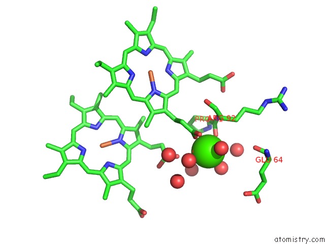
Mono view
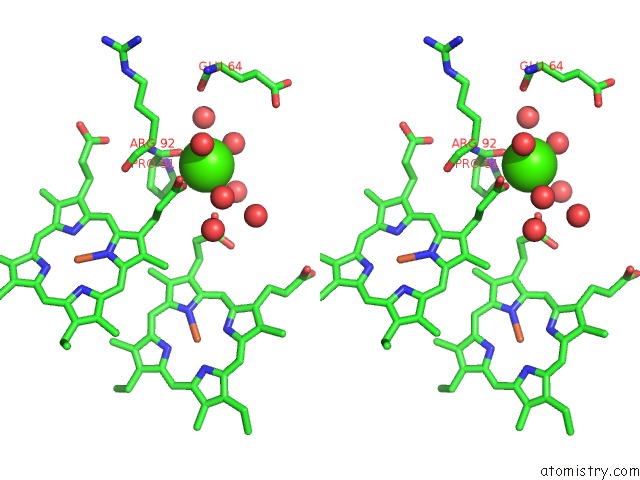
Stereo pair view
|
A full contact list of Calcium with other atoms in the Ca binding
site number 8 of High Resolution Crystal Structure of the Escherichia Coli Cytochrome C Nitrite Reductase. within 5.0Å range:
probe
|
atom
|
residue
|
distance (Å)
|
B
|
Occ
|
D:Ca1502
b:16.3
occ:0.50
|
O
|
D:PRO91
|
2.3
|
10.9
|
1.0
|
O1A
|
D:HEC3
|
2.3
|
12.1
|
1.0
|
O
|
D:HOH707
|
2.6
|
27.7
|
1.0
|
O
|
D:HOH758
|
2.8
|
36.2
|
1.0
|
C
|
D:PRO91
|
3.4
|
10.5
|
1.0
|
O1A
|
D:HEC4
|
3.4
|
26.5
|
1.0
|
CGA
|
D:HEC3
|
3.4
|
12.8
|
1.0
|
O2A
|
D:HEC3
|
3.8
|
10.8
|
1.0
|
O
|
D:HOH974
|
3.8
|
29.5
|
1.0
|
CA
|
D:ARG92
|
3.9
|
11.3
|
1.0
|
N
|
D:ARG92
|
4.1
|
10.7
|
1.0
|
O
|
D:HOH973
|
4.3
|
33.2
|
1.0
|
CA
|
D:PRO91
|
4.4
|
10.2
|
1.0
|
CB
|
D:ARG92
|
4.5
|
11.8
|
1.0
|
N
|
D:PRO91
|
4.6
|
10.3
|
1.0
|
CGA
|
D:HEC4
|
4.6
|
22.5
|
1.0
|
O
|
D:HOH776
|
4.6
|
32.5
|
1.0
|
CB
|
D:PRO91
|
4.7
|
10.8
|
1.0
|
O
|
D:HOH709
|
4.7
|
34.4
|
1.0
|
CBA
|
D:HEC3
|
4.7
|
9.0
|
1.0
|
CD
|
D:PRO91
|
4.8
|
11.0
|
1.0
|
O
|
D:HOH700
|
4.8
|
25.3
|
1.0
|
CAA
|
D:HEC3
|
4.8
|
8.2
|
1.0
|
C
|
D:ARG92
|
4.9
|
10.6
|
1.0
|
O
|
D:GLU64
|
5.0
|
17.0
|
1.0
|
|
Reference:
T.A.Clarke,
G.L.Kemp,
J.H.Wonderen,
R.M.Doyle,
J.A.Cole,
N.Tovell,
M.R.Cheesman,
J.N.Butt,
D.J.Richardson,
A.M.Hemmings.
Role of A Conserved Glutamine Residue in Tuning the Catalytic Activity of Escherichia Coli Cytochrome C Nitrite Reductase. Biochemistry V. 47 3789 2008.
ISSN: ISSN 0006-2960
PubMed: 18311941
DOI: 10.1021/BI702175W
Page generated: Fri Jul 12 15:52:01 2024
|