Calcium »
PDB 2rf7-2tep »
2sba »
Calcium in PDB 2sba: Soybean Agglutinin Complexed with 2,6-Pentasaccharide
Protein crystallography data
The structure of Soybean Agglutinin Complexed with 2,6-Pentasaccharide, PDB code: 2sba
was solved by
A.Dessen,
D.Gupta,
S.Sabesan,
C.F.Brewer,
J.C.Sacchettini,
with X-Ray Crystallography technique. A brief refinement statistics is given in the table below:
| Resolution Low / High (Å) | 90.00 / 2.60 |
| Space group | P 64 2 2 |
| Cell size a, b, c (Å), α, β, γ (°) | 144.900, 144.900, 109.400, 90.00, 90.00, 120.00 |
| R / Rfree (%) | n/a / n/a |
Other elements in 2sba:
The structure of Soybean Agglutinin Complexed with 2,6-Pentasaccharide also contains other interesting chemical elements:
| Manganese | (Mn) | 1 atom |
Calcium Binding Sites:
The binding sites of Calcium atom in the Soybean Agglutinin Complexed with 2,6-Pentasaccharide
(pdb code 2sba). This binding sites where shown within
5.0 Angstroms radius around Calcium atom.
In total only one binding site of Calcium was determined in the Soybean Agglutinin Complexed with 2,6-Pentasaccharide, PDB code: 2sba:
In total only one binding site of Calcium was determined in the Soybean Agglutinin Complexed with 2,6-Pentasaccharide, PDB code: 2sba:
Calcium binding site 1 out of 1 in 2sba
Go back to
Calcium binding site 1 out
of 1 in the Soybean Agglutinin Complexed with 2,6-Pentasaccharide
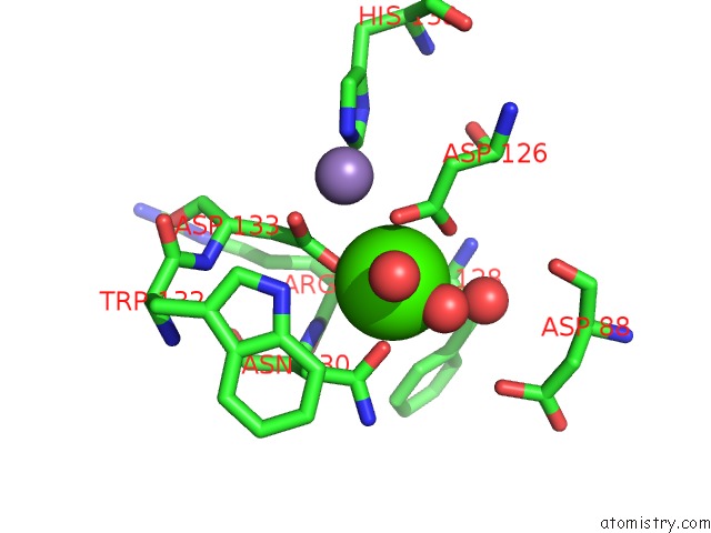
Mono view
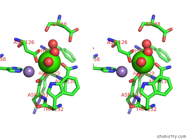
Stereo pair view
Mono view
Stereo pair view
A full contact list of Calcium with other atoms in the Ca binding
site number 1 of Soybean Agglutinin Complexed with 2,6-Pentasaccharide within 5.0Å range:
|
Reference:
A.Dessen,
D.Gupta,
S.Sabesan,
C.F.Brewer,
J.C.Sacchettini.
X-Ray Crystal Structure of the Soybean Agglutinin Cross-Linked with A Biantennary Analog of the Blood Group I Carbohydrate Antigen. Biochemistry V. 34 4933 1995.
ISSN: ISSN 0006-2960
PubMed: 7711015
DOI: 10.1021/BI00015A004
Page generated: Tue Jul 8 08:21:20 2025
ISSN: ISSN 0006-2960
PubMed: 7711015
DOI: 10.1021/BI00015A004
Last articles
Cl in 5FZ4Cl in 5FZ3
Cl in 5FYY
Cl in 5FYE
Cl in 5FYX
Cl in 5FYV
Cl in 5FYU
Cl in 5FYT
Cl in 5FYS
Cl in 5FWV