Calcium »
PDB 2tga-2v5c »
2trm »
Calcium in PDB 2trm: The Three-Dimensional Structure of ASN102 Mutant of Trypsin. Role of ASP102 in Serine Protease Catalysis
Enzymatic activity of The Three-Dimensional Structure of ASN102 Mutant of Trypsin. Role of ASP102 in Serine Protease Catalysis
All present enzymatic activity of The Three-Dimensional Structure of ASN102 Mutant of Trypsin. Role of ASP102 in Serine Protease Catalysis:
3.4.21.4;
3.4.21.4;
Protein crystallography data
The structure of The Three-Dimensional Structure of ASN102 Mutant of Trypsin. Role of ASP102 in Serine Protease Catalysis, PDB code: 2trm
was solved by
R.M.Stroud,
J.Finer-Moore,
with X-Ray Crystallography technique. A brief refinement statistics is given in the table below:
| Resolution Low / High (Å) | 7.00 / 2.80 |
| Space group | I 2 3 |
| Cell size a, b, c (Å), α, β, γ (°) | 124.380, 124.380, 124.380, 90.00, 90.00, 90.00 |
| R / Rfree (%) | n/a / n/a |
Calcium Binding Sites:
The binding sites of Calcium atom in the The Three-Dimensional Structure of ASN102 Mutant of Trypsin. Role of ASP102 in Serine Protease Catalysis
(pdb code 2trm). This binding sites where shown within
5.0 Angstroms radius around Calcium atom.
In total only one binding site of Calcium was determined in the The Three-Dimensional Structure of ASN102 Mutant of Trypsin. Role of ASP102 in Serine Protease Catalysis, PDB code: 2trm:
In total only one binding site of Calcium was determined in the The Three-Dimensional Structure of ASN102 Mutant of Trypsin. Role of ASP102 in Serine Protease Catalysis, PDB code: 2trm:
Calcium binding site 1 out of 1 in 2trm
Go back to
Calcium binding site 1 out
of 1 in the The Three-Dimensional Structure of ASN102 Mutant of Trypsin. Role of ASP102 in Serine Protease Catalysis
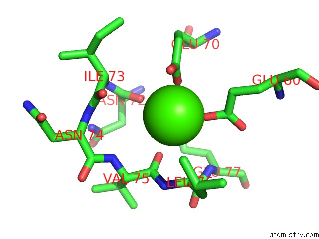
Mono view
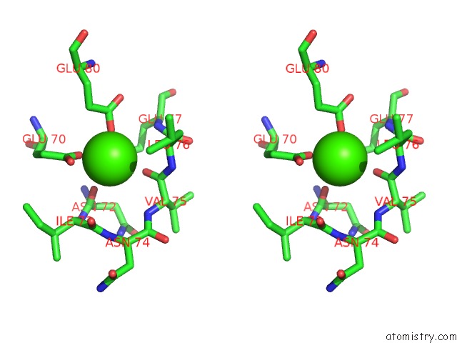
Stereo pair view
Mono view
Stereo pair view
A full contact list of Calcium with other atoms in the Ca binding
site number 1 of The Three-Dimensional Structure of ASN102 Mutant of Trypsin. Role of ASP102 in Serine Protease Catalysis within 5.0Å range:
|
Reference:
S.Sprang,
T.Standing,
R.J.Fletterick,
R.M.Stroud,
J.Finer-Moore,
N.H.Xuong,
R.Hamlin,
W.J.Rutter,
C.S.Craik.
The Three-Dimensional Structure of ASN102 Mutant of Trypsin: Role of ASP102 in Serine Protease Catalysis. Science V. 237 905 1987.
ISSN: ISSN 0036-8075
PubMed: 3112942
Page generated: Fri Jul 12 17:44:23 2024
ISSN: ISSN 0036-8075
PubMed: 3112942
Last articles
Zn in 9J0NZn in 9J0O
Zn in 9J0P
Zn in 9FJX
Zn in 9EKB
Zn in 9C0F
Zn in 9CAH
Zn in 9CH0
Zn in 9CH3
Zn in 9CH1