Calcium »
PDB 2tga-2v5c »
2uvw »
Calcium in PDB 2uvw: Crystal Structures of Mutant DPO4 Dna Polymerases with 8- Oxog Containing Dna Template-Primer Constructs
Enzymatic activity of Crystal Structures of Mutant DPO4 Dna Polymerases with 8- Oxog Containing Dna Template-Primer Constructs
All present enzymatic activity of Crystal Structures of Mutant DPO4 Dna Polymerases with 8- Oxog Containing Dna Template-Primer Constructs:
2.7.7.7;
2.7.7.7;
Protein crystallography data
The structure of Crystal Structures of Mutant DPO4 Dna Polymerases with 8- Oxog Containing Dna Template-Primer Constructs, PDB code: 2uvw
was solved by
A.Irimia,
M.Egli,
with X-Ray Crystallography technique. A brief refinement statistics is given in the table below:
| Resolution Low / High (Å) | 30.81 / 2.09 |
| Space group | P 21 21 2 |
| Cell size a, b, c (Å), α, β, γ (°) | 96.778, 103.728, 52.915, 90.00, 90.00, 90.00 |
| R / Rfree (%) | 24.9 / 27.4 |
Calcium Binding Sites:
The binding sites of Calcium atom in the Crystal Structures of Mutant DPO4 Dna Polymerases with 8- Oxog Containing Dna Template-Primer Constructs
(pdb code 2uvw). This binding sites where shown within
5.0 Angstroms radius around Calcium atom.
In total 3 binding sites of Calcium where determined in the Crystal Structures of Mutant DPO4 Dna Polymerases with 8- Oxog Containing Dna Template-Primer Constructs, PDB code: 2uvw:
Jump to Calcium binding site number: 1; 2; 3;
In total 3 binding sites of Calcium where determined in the Crystal Structures of Mutant DPO4 Dna Polymerases with 8- Oxog Containing Dna Template-Primer Constructs, PDB code: 2uvw:
Jump to Calcium binding site number: 1; 2; 3;
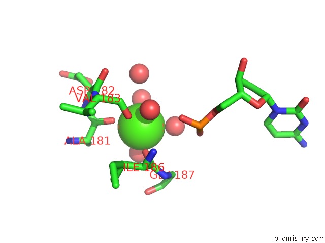
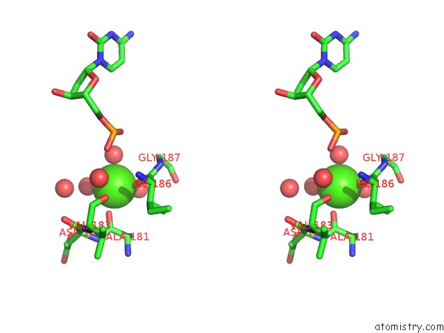
Calcium binding site 1 out of 3 in 2uvw
Go back to
Calcium binding site 1 out
of 3 in the Crystal Structures of Mutant DPO4 Dna Polymerases with 8- Oxog Containing Dna Template-Primer Constructs
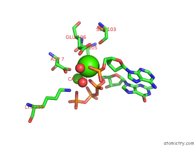
Mono view
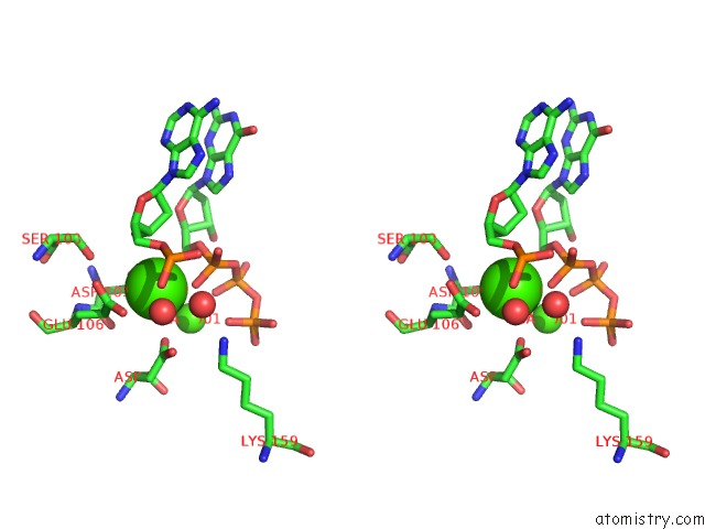
Stereo pair view
Mono view
Stereo pair view
A full contact list of Calcium with other atoms in the Ca binding
site number 1 of Crystal Structures of Mutant DPO4 Dna Polymerases with 8- Oxog Containing Dna Template-Primer Constructs within 5.0Å range:
|
Calcium binding site 2 out of 3 in 2uvw
Go back to
Calcium binding site 2 out
of 3 in the Crystal Structures of Mutant DPO4 Dna Polymerases with 8- Oxog Containing Dna Template-Primer Constructs
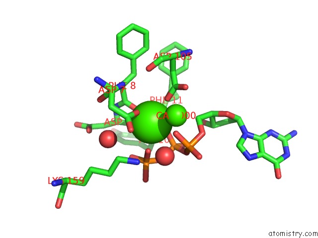
Mono view
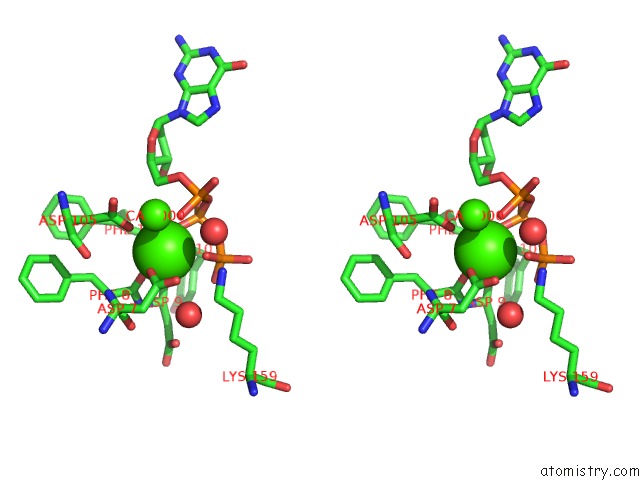
Stereo pair view
Mono view
Stereo pair view
A full contact list of Calcium with other atoms in the Ca binding
site number 2 of Crystal Structures of Mutant DPO4 Dna Polymerases with 8- Oxog Containing Dna Template-Primer Constructs within 5.0Å range:
|
Calcium binding site 3 out of 3 in 2uvw
Go back to
Calcium binding site 3 out
of 3 in the Crystal Structures of Mutant DPO4 Dna Polymerases with 8- Oxog Containing Dna Template-Primer Constructs
Mono view
Stereo pair view
Mono view
Stereo pair view
A full contact list of Calcium with other atoms in the Ca binding
site number 3 of Crystal Structures of Mutant DPO4 Dna Polymerases with 8- Oxog Containing Dna Template-Primer Constructs within 5.0Å range:
|
Reference:
R.L.Eoff,
A.Irimia,
K.C.Angel,
M.Egli,
F.P.Guengerich.
Hydrogen Bonding of 7,8-Dihydro-8- Oxodeoxyguanosine with A Charged Residue in the Little Finger Domain Determines Miscoding Events in Sulfolobus Solfataricus Dna Polymerase DPO4. J.Biol.Chem. V. 282 19831 2007.
ISSN: ISSN 0021-9258
PubMed: 17468100
DOI: 10.1074/JBC.M702290200
Page generated: Tue Jul 8 08:27:19 2025
ISSN: ISSN 0021-9258
PubMed: 17468100
DOI: 10.1074/JBC.M702290200
Last articles
Fe in 2YXOFe in 2YRS
Fe in 2YXC
Fe in 2YNM
Fe in 2YVJ
Fe in 2YP1
Fe in 2YU2
Fe in 2YU1
Fe in 2YQB
Fe in 2YOO