Calcium »
PDB 2tga-2v5c »
2ux1 »
Calcium in PDB 2ux1: Identification of Two Zinc-Binding Sites in the Streptococcus Suis Dpr Protein
Protein crystallography data
The structure of Identification of Two Zinc-Binding Sites in the Streptococcus Suis Dpr Protein, PDB code: 2ux1
was solved by
H.Havukainen,
A.Kauko,
A.T.Pulliainen,
S.Haataja,
W.Meyer-Klaucke,
J.Finne,
A.C.Papageorgiou,
with X-Ray Crystallography technique. A brief refinement statistics is given in the table below:
| Resolution Low / High (Å) | 19.97 / 1.80 |
| Space group | P 21 21 21 |
| Cell size a, b, c (Å), α, β, γ (°) | 104.520, 137.690, 142.060, 90.00, 90.00, 90.00 |
| R / Rfree (%) | 20.6 / 24.8 |
Other elements in 2ux1:
The structure of Identification of Two Zinc-Binding Sites in the Streptococcus Suis Dpr Protein also contains other interesting chemical elements:
| Chlorine | (Cl) | 2 atoms |
| Zinc | (Zn) | 24 atoms |
Calcium Binding Sites:
The binding sites of Calcium atom in the Identification of Two Zinc-Binding Sites in the Streptococcus Suis Dpr Protein
(pdb code 2ux1). This binding sites where shown within
5.0 Angstroms radius around Calcium atom.
In total 4 binding sites of Calcium where determined in the Identification of Two Zinc-Binding Sites in the Streptococcus Suis Dpr Protein, PDB code: 2ux1:
Jump to Calcium binding site number: 1; 2; 3; 4;
In total 4 binding sites of Calcium where determined in the Identification of Two Zinc-Binding Sites in the Streptococcus Suis Dpr Protein, PDB code: 2ux1:
Jump to Calcium binding site number: 1; 2; 3; 4;
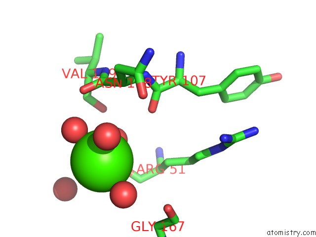
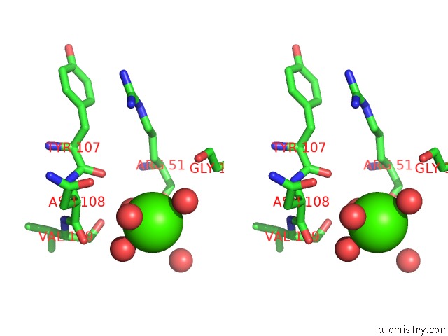
Calcium binding site 1 out of 4 in 2ux1
Go back to
Calcium binding site 1 out
of 4 in the Identification of Two Zinc-Binding Sites in the Streptococcus Suis Dpr Protein

Mono view

Stereo pair view
Mono view
Stereo pair view
A full contact list of Calcium with other atoms in the Ca binding
site number 1 of Identification of Two Zinc-Binding Sites in the Streptococcus Suis Dpr Protein within 5.0Å range:
|
Calcium binding site 2 out of 4 in 2ux1
Go back to
Calcium binding site 2 out
of 4 in the Identification of Two Zinc-Binding Sites in the Streptococcus Suis Dpr Protein

Mono view

Stereo pair view
Mono view
Stereo pair view
A full contact list of Calcium with other atoms in the Ca binding
site number 2 of Identification of Two Zinc-Binding Sites in the Streptococcus Suis Dpr Protein within 5.0Å range:
|
Calcium binding site 3 out of 4 in 2ux1
Go back to
Calcium binding site 3 out
of 4 in the Identification of Two Zinc-Binding Sites in the Streptococcus Suis Dpr Protein
Mono view
Stereo pair view
Mono view
Stereo pair view
A full contact list of Calcium with other atoms in the Ca binding
site number 3 of Identification of Two Zinc-Binding Sites in the Streptococcus Suis Dpr Protein within 5.0Å range:
|
Calcium binding site 4 out of 4 in 2ux1
Go back to
Calcium binding site 4 out
of 4 in the Identification of Two Zinc-Binding Sites in the Streptococcus Suis Dpr Protein

Mono view

Stereo pair view
Mono view
Stereo pair view
A full contact list of Calcium with other atoms in the Ca binding
site number 4 of Identification of Two Zinc-Binding Sites in the Streptococcus Suis Dpr Protein within 5.0Å range:
|
Reference:
H.Havukainen,
S.Haataja,
A.Kauko,
A.T.Pulliainen,
A.Salminen,
T.Haikarainen,
J.Finne,
A.C.Papageorgiou.
Structural Basis of the Zinc- and Terbium-Mediated Inhibition of Ferroxidase Activity in Dps Ferritin- Like Proteins. Protein Sci. V. 17 1513 2008.
ISSN: ISSN 0961-8368
PubMed: 18552126
DOI: 10.1110/PS.036236.108
Page generated: Tue Jul 8 08:27:40 2025
ISSN: ISSN 0961-8368
PubMed: 18552126
DOI: 10.1110/PS.036236.108
Last articles
Cl in 5R9MCl in 5R9K
Cl in 5R9L
Cl in 5R9J
Cl in 5R9I
Cl in 5R9G
Cl in 5R9H
Cl in 5R9F
Cl in 5R9C
Cl in 5R9E